
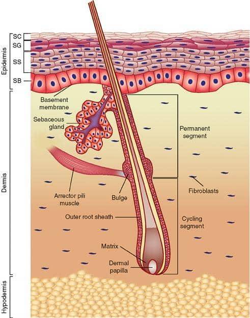
The pavement cells They are lining epithelial cells with large nuclei and large amounts of cytoplasm. These cells are present in almost all tissues of eukaryotic organisms. In animals, squamous cells form the lining epithelial tissue that lines the outer body surface, internal organs and ducts..
Pavement cells are easy to identify under the microscope using silver nitrate, as they appear with a typical ordered mosaic appearance composed of hexagonal cells with irregular contours..
Typical pavement cells have a very thin and elongated cytoplasm, distributed longitudinally with a central bulge where the nucleus is located. These cells have the appearance of a spaceship or flying saucer..
The skin is made up almost entirely of paving cells, where they perform protection functions, increase the number of cells, secretion and perception and detection of external stimuli..
Article index
Paving cells are classified into three types according to the anatomical area they occupy, their topological and morphological characteristics. The three known types of pavement cells are:
-Flat paving cells: they are elongated with large nuclei. Found in blood and lymphatic vessels, kidney, heart, and lungs.
-Cubic paving cells: They have a large amount of cytoplasm and are involved in the secretory functions of tissues. These line the ovaries, oral cavity, esophagus, anus, and some areas of the brain..
-Prismatic paving cells: they are found in the basal laminae of the tissue, they may have cilia to facilitate transport. These cells make up almost all the glands in the body..
In animals, squamous cells are part of monostratified, pseudostratified, and multilayer epithelial tissue.
In monostratified epithelial tissue, squamous cells form a thin layer organized in rows of cells, this being the most superficial portion of the tissue..
The pseudostratified tissue is composed exclusively of a single layer of squamous epithelial cells, which are found in a disorderly manner.
Pavement cells in polylayer epithelial tissue are stacked in layers of axially elongated cells, almost completely flat. In this epithelium the cells are closely adhered to each other and arranged in several layers on the basement membrane..
Paving cells act as a protective barrier that prevents the entry of pathogenic microorganisms into our body. These cells are part of our primary immune system, protecting us from external aggressions and mechanical trauma..
Paving cells regulate the degree of hydration and the loss of water by evaporation. In the serous cavities, the lining with these cells facilitates the movement of the viscera and that of the food..
In the endothelium of blood vessels, squamous cells allow the diffusion of water and ions by active transport (pinocytosis), and at the same time prevent the entry of macromolecules into the tissue..
In women, squamous cells are part of the cervix, vagina, vulva, and vaginal secretions. The gynecological study of these cells is of great informative value to know the health of the reproductive organ.
Some of these cells are endowed with nerve endings and fulfill an important sensory function in the reproductive organs..
In organisms such as teleost fish (trout), it has been proposed that squamous cells are directly involved in the ionic transport of sodium, which is actively diffused by flat squamous cells.
Pavement cell screening is a common technique for finding vesicular skin pathologies in stratified epithelium. Squamous cells with secretory functions are highly susceptible to viral and bacterial infections.
In women, the squamous cells shed in a cyclical way, depending on the variable hormonal levels and according to the stage of the organism's life cycle.
It is customary to study vaginal squamous cells using the Papanicolaou staining method, introduced by Dr. G. N. Papanicolaou in 1942. This method links cell type morphology with endocrinology and histology.
Cytological studies of the squamous epithelial cells of the uterine area allow determining if there is the presence of the Human Papilloma Virus (HPV).
The identification of the morphological changes in the squamous cells provides useful information for the cytodiagnosis of cancer, allowing to differentiate preneoplastic and neoplastic alterations.
Paving cells can present mild alterations, benign abnormalities, inflammatory and reactive changes. These alterations can be the product of the normal behavior of the organism or they can be related to pathological disorders and relevant diseases..
Paving cells have normal phenotypic growths and masses mediated by hormones, which modify their texture, degree of secretion, and metabolism. These changes may be typical of tissue aging.
Benign abnormalities may include mild inflammation, an increase or decrease in the number of epithelial squamous cells, and rarely scarification or keratinization of the epithelial cells..
Inflammatory abnormalities in squamous cells are identified in the nucleus, which implies a decrease or loss of cellular activity. This decrease in cell activity typically leads to cell death from necrosis..
Typical inflammatory abnormalities include:
There are inflammatory abnormalities that are directly related to certain pathologies. Among these are the presence of deep cells and Atrophic Colpitis or vaginitis.
Deep cells in women of childbearing age are normal, as they are the product of menstrual cycles that exfoliate the squamous cells of the cervix and vagina. However, its existence in infants and elderly women is related to diseases.
These diseases include some severe inflammatory reactions in the cervix and vagina, damage to the reproductive system, hormonal imbalances, or the presence of pathogenic agents..
Atrophic Colpitis is produced by the disappearance of layers of pavenous cells during differentiation, reducing the epithelia to a few rows of parabasal cells..
The reduction in the differentiation of epithelia is the product of hypoestrogenism, since this stops the mechanisms of cell division and differentiation.
Reactive changes are generally benign and are associated with abnormalities that clinicians cannot accurately define on cytology examinations. However, these changes can appear when there are infections or other irritations.


Yet No Comments