The satellite cells they are skeletal muscle cells. They are small, uninucleated cells that are in a quiescent (dormant) state in adult mammals, which is why they are said to function as a population of "reserve" cells capable of proliferating under certain conditions..
The skeletal muscle of mammalian animals and many other vertebrates is made up of muscle cells, also called muscle fibers, which are the completely differentiated cells that contain the contractile elements or proteins of this tissue..
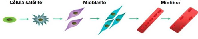
These muscle fibers are formed during development thanks to the migration of precursor muscle cells (myoblasts) from the embryonic "somites" to the nascent muscles, where they fuse with each other and form multinucleated muscle cells or myofibers (with more than one nucleus ).
In adult animals, muscle is formed or, rather, it regenerates, thanks to the proliferation of satellite cells, which were discovered in 1961 by A. Mauro. These cells are separated from the muscle fibers, as they are found under the basal lamina of each one..
It is a very important type of cell for mammalian muscle tissue, as these probably represent the only cellular source for muscle regeneration in adulthood, whether due to injury, damage, disease or physical exercise..
Although the term "satellite cell" is also used to distinguish a group of glial cells of the peripheral nervous system, which are located specifically in the sensory, sympathetic, and parasympathetic ganglia, it is more commonly used to refer to proliferating muscle cells that are newly did we mention.
Article index
Satellite cells are formed in the extremities during embryonic development, after the formation of the first muscle fibers (myofibers). These cells are closely associated with the plasma membrane of muscle cells (sarcolemma), as they reside between it and its basal lamina.
They are easily distinguishable due to their location and morphology, although they are very heterogeneous cell populations, that is, with very different cells.
This heterogeneity is based not only on their asymmetric division, but also on the expression of different proteins and transcription factors, on their organization, etc..
Muscle satellite cells can be molecularly distinguished from other cells thanks to the concomitant expression of different molecular markers, among which the transcription factors of the Pax family stand out..
Belonging to this family is the transcription factor Pax7, which apparently is essential for the maintenance of the "undifferentiated" state of satellite cells, as well as their capacity for self-renewal..
These cells also express the factor Pax3, which is extremely important during the initial steps of muscle formation and is involved in regulating the transcription of another marker known as the receptor tyrosine kinase c-Met..
In addition to Pax factors, satellite cells are known to co-express (express at the same time):
- The myogenesis (muscle building) regulatory factor known as Myf5
- The transcription factor Barx2, regulator of muscle growth, maintenance and regeneration
- The protein M-cadherin, a cell adhesion protein
- Integrin-7 surface binding receptor
- The differentiation group 34 protein, CD34
- The proteoglycans syndecane-3 and syndecane-4
- The CXCR4 chemokine receptor
- The caveolae-forming protein, caveolin-1
- A calcitonin receptor
- Vascular adhesion protein 1, VCAM-1
- The neural cell adhesion molecule 1, NCAM-1
- The nuclear envelope proteins Laminin A, Laminin C, and Emerin
The regenerative characteristics of muscle tissue are mainly due to the action of satellite cells, which function as a "reservoir" of precursor cells, responsible for postnatal growth and muscle regeneration after injury, physical exercise or the product of a disease..
When these cells proliferate, they usually do so asymmetrically, since a part of their progeny fuses with the growing muscle fibers and another is in charge of maintaining the population of regenerative satellite cells..
They are extremely abundant cells during muscle growth, but their number decreases with age.
Numerous experimental reports suggest that satellite cells are activated (come out of their normal quiescent state) when skeletal muscle suffers some damage or after heavy physical exercise..
This "activation" occurs by different signaling pathways and, once active, these cells proliferate and can do two things: (1) fuse with each other to form "myotubes" that mature to form myofibers or (2) fuse with the segments damaged existing muscle fibers (using them as "scaffolds" or "casts").
For this reason, these cells are also considered a kind of muscle “stem cells”, since they are capable of forming new muscle cells and of regenerating the population of satellite cells in the muscle that suffered some unforeseen.
For many authors, muscle regeneration mediated by satellite cells consists of a series of "steps" that closely resemble the phases of embryonic muscle development..
- Initially, satellite cells have to "come out" of their quiescent or dormant state and become activated, so that they can begin to divide..
- The division process, as discussed above, is asymmetric, which is necessary for some cells to commit to the formation of new muscle cells and others to maintain the "constant" number of quiescent cells..
- Thus, myoblasts, that is, the cells produced by satellite cells to regenerate muscle, fuse and form "myotubes". The myotubes can, in turn, fuse with each other or with a pre-existing fiber to repair it, which will later grow and mature.
The quiescence of the satellite cells must be maintained during the life of the muscle fibers, since these must be activated only when the appropriate signals indicate it..
Some experimental results suggest that, compared to active cells, quiescent satellite cells express 500 more genes, the products of which are surely involved in quiescence..


Yet No Comments