
A coccobacillus it is a bacterium with a cellular morphology intermediate between a coconut and a bacillus. It is usual for bacteria to be classified according to their cell shape, but many times the limits between these categories are not well established, an example of which is the coccobacilli.
A coconut is a spheroid-shaped bacterium, while the cells of the bacilli are more elongated and reminiscent of a rod. In the case of coccobacilli, the shape of the cell is a rod so short that it can easily be mistaken for a coconut..
There are a number of biological entities that exhibit coccobacilli morphology and are medically important..
Article index
Within prokaryotes, eubacteria exhibit enormous morphological diversity that allows the grouping of these organisms.
In the world of bacteria, the most common forms are: spherical-shaped cocci, bacilli that are straight cylinders of variable length similar to rods, and spirilli that are elongated corkscrews..
Of these three main forms, we find various variants and combinations. Among these modifications are vibrios, comma-shaped cells; corynebacteria, rods with a rounded end; and the coccobacilli, a short cane with an oval outline.
The morphological distinction does not provide additional information on the biology of the organism. In other words, knowing that a bacterium is a coccobacillus does not say anything about its structural and biochemical characteristics, among others..
Among the pathogens that exhibit a cocobacillus morphology we have the following prokaryotic species:
H. influenzae it is a coccobacillus that does not have structures that allow its mobility. Their metabolism is generally aerobic, but if environmental conditions warrant it, they can behave like anaerobic organisms. This metabolic tendency is called facultative anaerobic.
From a medical point of view, H. influenzae It has been linked to a wide range of diseases, from meningitis, pneumonia and sepsis, to other less severe diseases.
One of the most common ways of referring to bacteria is according to their response to the Gram stain. The coloration seeks to separate the bacteria according to the structure of their bacterial wall. This species is Gram negative.
Gram negative bacteria have a double cell membrane. Between them there is a small layer of peptidoglycan. Gram positive ones, on the other hand, are bacteria with a single plasma membrane, and above this a thick layer of peptidoglycan is located. This stain is very useful in microbiology.
G. vaginalis It is a bacterium that lives in the vagina of the human species. It does not have structures to move, so it is not mobile, it is facultative anaerobic (like the previous species), and it does not have the ability to form endospores.
It is related to bacterial vaginosis. The presence of this bacterium destabilizes the natural microbiota of the vagina, increasing the frequency of some genders and decreasing those of others..
The disease is usually asymptomatic, although the secretions are characteristic and have unpleasant odors. It can be transmitted sexually, although it is not considered a venereal disease. Many times the bacteria can remain on the female genitalia harmlessly.
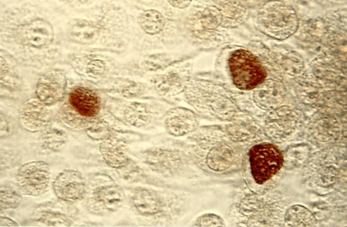
Bacteria of the species C. trachomatis are obligate pathogens that exclusively infect the human species and is the causative agent of chlamydia - a sexually prevalent disease of significant prevalence in human populations, affecting both men and women.
The bacteria can lodge in the cervix, urethra, rectum, or throat. Associated symptoms include pain in the genitals, burning when urinating, and abnormal secretions from the sexual organs..
Like the two bacteria we have described, A. actinomycetemcomitans it is an immobile bacterium. Responds negatively when Gram stain is applied.
It has been associated with the generation of an oral disease called periodontitis. Patients who suffer from this condition present loss of collagen and if it is not treated it can lead to extreme consequences such as bone loss, leaving the tooth without bone support..
The probability of acquiring the disease is increased by other conditions such as diabetes or certain imbalances of the immune system, in addition to unhealthy lifestyle habits such as smoking.
The morphology of the bacteria usually changes depending on the conditions. When grown in the laboratory, cells more closely resemble a rod - an average bacillus. But, when looking at the direct shapes live, the shape is more spherical, like a coconut..
The elimination of the bacteria can be done with the taking of antibiotics. In extreme cases, healthcare professionals resort to surgical removal.
B. pertussis are organisms that live strictly in aerobic environments, are immobile and respond negatively to the Gram stain.
It is the cause of the condition called whooping cough or whooping cough that exclusively affects humans. The infection is extremely contagious and occurs through violent coughing and choking sensations..
Together, the patient has trachebronchial inflation. As the infection progresses, complications spread to other systems, compromising organs of the nervous system and the circulatory system. The prevalence is higher in developing countries and in infants under five years of age.
However, recently (in 2010 and 2012) two outbreaks of pertussis have been reported in different regions of the United States..
Bacteria of the same genus are associated with coughing episodes in humans, but they are milder pathologies.
Y. pestis It is a facultative anaerobic enterobacterium that responds negatively to Gram stain. It is the agent of different infections that affect humans, including pulmonary plague, bubonic plague and, to a lesser extent, septicemic plague..
Historically, the consequences of the prevalence of the disease have been devastating for human populations, being the cause of multiple pandemics. In fact, it has caused more deaths than any other infectious disease, second only to malaria..


Yet No Comments