
A surgical drainage it is a medical method of removing blood, pus, or other fluids from an operative site. It can be placed in an abscess, for example, to speed recovery from a localized infection, or in a cyst or seroma, to remove fluids and cells. Drains can also be inserted into clogged organs to relieve pressure resulting from fluid buildup within the organs..
Drains remove blood, serum, lymph, and other fluids that collect in the wound bed after a procedure. If allowed to develop, these fluids put pressure on the surgical site, as well as on adjacent organs, vessels, and nerves..
Decreased perfusion delays healing; increased pressure causes pain. Additionally, a buildup of fluid serves as a breeding ground for bacteria. Fluid can be removed from a wound using passive or active surgical drainage.
Passive drains rely on gravity to evacuate fluid, while active drains are attached to a vacuum or suction device in the wall. A surgeon chooses a drain that fits both the operative site and can handle the type and amount of drainage expected..
For example, a T-tube is a fairly large passive drain that is typically placed during a cholecystectomy to accommodate the 200 - 500 ml of bile that is expected to accumulate in the early postoperative period..
The Penrose is another passive drain that is generally placed to handle smaller amounts of drain. That's a good thing, because it is normally left open, that is, its free end, which protrudes an inch above the skin, is not usually connected to a bag to collect drainage.
Instead, fluid from the wounds seeps out onto a gauze pad. Active drains like the Jackson-Pratt (JP) and Hemovac always have a drain pan. Drains that have some type of bag are often called closed systems.
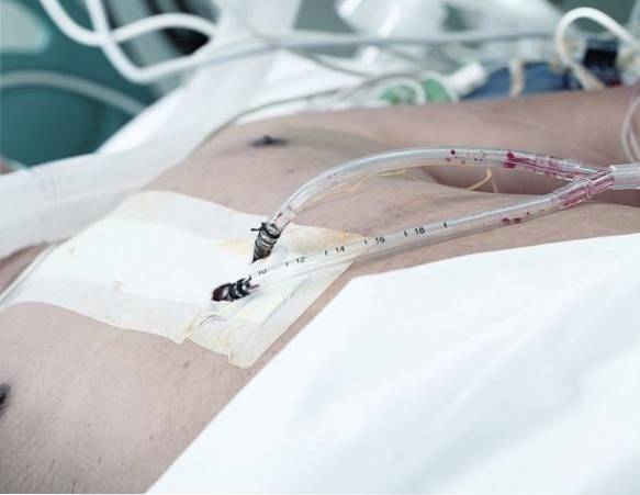
Unlike the Penrose, the ducts in a JP or Hemovac are slightly stiffer so they won't flatten under the pressure exerted by suction. The tips of these drains are fenestrated, which means they have multiple holes to facilitate drainage. In either case, a drain can come out of a wound through the suture line or through a small opening near the incision..
Article index
Drains can be:
Open drains (including corrugated rubber or plastic sheeting) drain fluid into a gauze pad or stoma bag. They are likely to increase the risk of infection.
Closed drains are made up of tubes that drain into a bag or bottle. Examples include chest, abdominal, and orthopedic drains. In general, the risk of infection is reduced.
Active drains are kept under suction (which can be low or high pressure). Passive drains have no suction and work according to the differential pressure between the body cavities and the exterior.
They are relatively inert that induce minimal tissue reaction and rubbery drains that can induce a strong tissue reaction, sometimes allowing a tract to form (this can be considered useful, for example, with bile T tubes).
The downside to a drain is that it can be painful going in and out. Depending on the case, it can be painful to just sit on the wound. That's because the drainage destroys the tissue..
A drain also provides a path for bacteria to enter the wound. In fact, the risk of infection from a drain increases significantly on the third or fourth postoperative day, as does the degree of mechanical damage to local tissue.
To minimize these problems, the surgeon will place a drain so that it reaches the skin by the shortest and safest route. In this way, the drain exerts the least amount of pressure on the adjacent tissue..
However, to be effective, a drain must also reach the deepest and most dependent area of a wound to adequately evacuate excess fluid..
Unfortunately, the deeper a drain, the greater the risk of complications. And because the drainage is strange, the body quickly begins to close it in a granulation tissue..
Surgical drains are used in a wide variety of surgeries. Generally speaking, the intention is to decompress or drain fluid or air from the surgery area..
Examples:
Management is governed by the type, purpose, and location of the drain. It is common for the surgeon's preferences and instructions to be followed. A written protocol can assist ward staff with aftercare for surgical drains.
If active, the drain can be connected to a suction source (and adjusted to a prescribed pressure). It must be certified that the drainage is secure (detachment is likely to occur when transferring patients after anesthesia).
The shedding can increase the risk of infection and irritation of the surrounding skin. Drainage production must be accurately measured and recorded.
Changes in the character or volume of the fluid should be monitored and any complications that result in fluid leakage (particularly bile or pancreatic secretions) or blood should be identified. Fluid loss measurements should be used to aid intravenous fluid replacement.
In general, drains should be removed once drainage has stopped or becomes less than about 25 ml / day. Drains can be "shortened" by gradually withdrawing them (typically 2 cm per day) and thus, in theory, allowing the site to heal gradually.
Generally, the drains that protect postoperative sites from leakage form a tract and stay in place longer (usually for a week).
The patient should be advised that there may be some discomfort when the drain is removed. Early removal of the drain can decrease the risk of some complications, especially infection..

Yet No Comments