
The eyes are the two organs through which we can see everything that surrounds us, that is, they are the ones that allow us to have the sense of vision that, together with touch, hearing, smell and taste is one of the 5 senses; vision is a very complex process that depends on the different parts of our eyes.
Although the eyes may seem small in relation to the rest of our body, the eyes, that pair of small moving cameras that we have in the upper front part of our face, are two very interesting and complex organs. Other animals have them too, some very similar to ours and others more or less developed.
The eyes allow us to obtain visual information of what surrounds us and also determine some interesting characteristics of what we see, such as colors, shapes, the relative distance that we are from an object, the size and depth of the space where we are, among other things.
The eyes also have their own protection system, as they are capable of producing tears that lubricate and clean them when necessary..
Article index
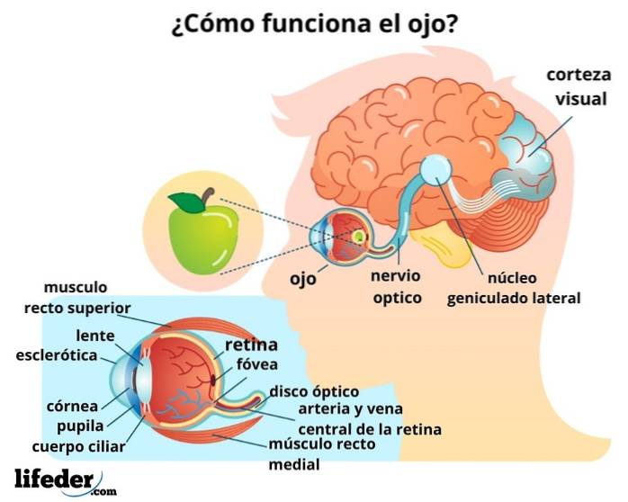
The sense of vision takes place when light "collides" with objects and the reflections of this light are irradiated towards the eyes that, when received, convert it into chemical or electrical information that is understandable to our brain, that is, into impulses. nervous that our brain interprets as images.
Our eyes work constantly during the day, while they are open when we are awake, and they rest when we go to sleep and close them..

Normally we all have two eyes, one next to the other, separated by the nose. Each of our eyes is about the size of a ping-pong ball, so they are not too big, but they are not too tiny.
The eyes are perfectly positioned in two identical hollow cavities that are in our skull: the eye cavities.
These cavities are formed by a region of our skull known as eye orbit. This cavity has a shape similar to that of a pyramid whose pointed end is directed towards the inside of the head and whose base "opens" towards the outside of the skull..
The orbit of each of our eyes is made up of a series of special bones, these are their names: frontal, sphenoid, zygomatic, maxillary, ethmoid, lacrimal and palatal.
Our two eyes are perfectly positioned in their eye sockets thanks to the fact that they are connected to a series of muscles called extraocular muscles. These muscles not only hold them in place, but allow us to move them in many different directions when looking at something..
There are 6 extraocular muscles and scholars of human anatomy divide them into two groups according to the type of movement they facilitate: the rectus muscles and the oblique muscles..
There are 4 rectus muscles: the superior rectus, the inferior rectus, the medial rectus and the medial lateral. There are two oblique muscles: one upper and one lower.
Above the eyes are the eyelids, which are portions of tissue that form the front of these and whose main function is to protect them from excessive light, airborne particles or any dangerous object, to clean them and keep them permanently moist through the "blink", which is a process voluntary and involuntary at the same time.
Both the inner part of the eyelids and the surface of the eyes are covered by a transparent mucous membrane called conjunctiva. This delicate membrane protects the eyeballs and participates in the formation of tears, as well as in the immune defense of the eyes..
Our eyes have a set of glands capable of producing substances that we call tears, which constantly lubricate and protect them. Tears are made up of three different elements: one watery, another oily (oily) and another mucous.
The watery part of tears is produced by the lacrimal glands, which are located internally under our eyebrows, in the region furthest from the nose..
The oily part, on the other hand, is produced by the meibomian glands, located in both the upper and lower eyelids. Finally, the mucous part is produced by the conjunctival membrane.
In addition to everything we have named, the eyes have other parts, all very different from each other, let's see what they are:
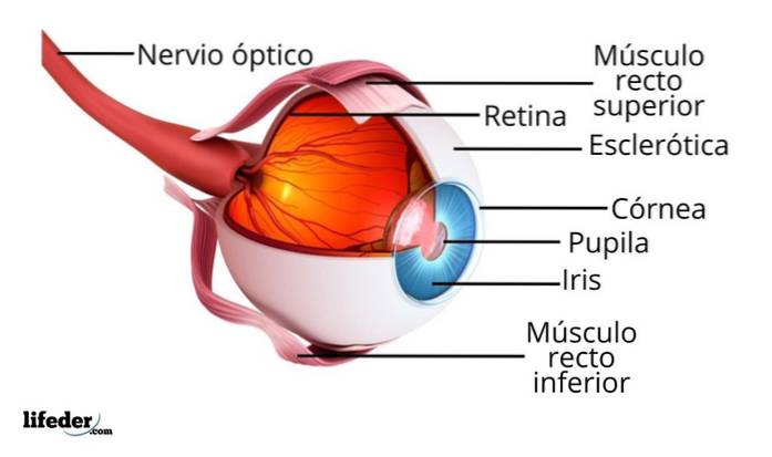
The whites of the eyes are known as the sclera. This region is formed by a very resistant tissue and its function is to cover most of the eyeball, which is what we will call the eye from now on, without taking into account the eyelids..
In the sclera we can find blood capillaries, which are small pipes whose main function is to irrigate blood to the cells of the eye, feeding them and providing everything they need to function properly..
If we stare at ourselves in the mirror or look at the eyes of another person, we can see that there is a white portion that surrounds the region that has color, that is the sclera.
In front of the colored portion of our eyes, which we will talk about immediately, there is a kind of transparent “dome” called “cornea”. The cornea is responsible for helping the eye to focus the light it receives when we are looking at something.
The cornea is made up of a transparent tissue, so it looks like glass, as if it were the window that shows the eye everything that is around us.
Between the cornea and the iris there is a small space known as the "anterior chamber" and it contains a transparent liquid responsible for nourishing our eyes and keeping them healthy..
The liquid contained in the anterior chamber is what is known as aqueous humor, which is constantly produced by the eyes. If necessary, this liquid can be drained, especially when the pressure inside the chamber rises dangerously..
The colored part of our eyes, the one by which we say that someone has brown, green, blue, black or gray eyes, is called “iris”. The iris is located just behind the cornea, that is, it is protected by this.
This region of our eyes is associated with very delicate muscles that help it to change shape depending on the need, as this serves to control the amount of light that passes into the pupil..
The pupil is the part of the eye that we see as a black point in the center of the iris (an opening of the iris) and it is the one that enlarges or shrinks due to the contraction or relaxation of the iris muscles, which are responsible for controlling the amount of light that passes.
When we are in very bright places, the pupil looks like a small black dot and it seems that the iris is much larger, since it responds to changes in the intensity of light.
On the other hand, if we have to strain our eyes to be able to look in the dark, the pupil is enlarged due to the absence of light, all in order to allow as much light as possible to enter the eye..
The parts of the eye that we have named so far are easily distinguishable with the naked eye, just by looking in a mirror or by looking closely at the eyes of another person.
Now, the internal portions of the eye can only be observed by specialists who have special devices for this purpose..
Such is the case of the crystalline lens, also called "lens", which is a transparent region located at the back of the iris and which works by focusing light rays towards the deepest region of the eyeball, which is known as the retina.
The lens or crystalline lens is suspended by muscle fibers that allow it to constantly change shape, which is necessary when we see things very close or very far..
The retina is at the back of the eye and receives light that has been focused and directed by the lens..
This region of our eyes has millions of cells that are sensitive to light and that are capable of converting light information in the form of nerve impulses, so that when these are transmitted to the brain, it can understand that we are seeing something.
Cells in the retina that are sensitive to light are called cones Y Canes. Canes help us see in black, white, and shades of gray; they also help us determine the shape of things. Cones, on the other hand, help us identify colors and color ranges.
The retina has a small, specialized region called taint, which is responsible for the central vision. Helps us get fine details of what we see, as well as things in motion.
Between the sclera and the retina is an additional layer of tissue called choroid membrane. It is a very thin and vascularized layer that contributes to the nutrition and oxygenation of the outermost cell layers of the retina. This membrane reflects light and is what causes the effect of "red eyes" in photographs..
The largest part of the eye is behind the lens and is known as the "vitreous body." We say that it is the largest part because it represents two thirds of the volume of our eyes and, therefore, it is who defines its shape.
Inside this body is contained a rather gelatinous fluid called the vitreous humor. When we see something, after the light passes through the lens, it goes directly into the vitreous humor at the back of our eyes..
Cells in the retina send special nerve messages to our brain, which give the brain information about what we see. These messages travel to the brain through the optic nerve, which is like a direct telephone line between the eyes and the brain..
Yet No Comments