The cerebral embolism, also known as embolic stroke, it is a type of cerebrovascular accident, that is, a temporary or permanent alteration of blood flow in one or more brain areas.
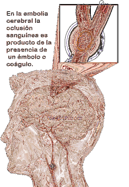
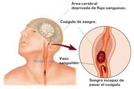
In cerebral embolism, blood occlusion is the product of the presence of an embolus, a body of organic matter (blood, fat or gas clot) that is located in an encephalic blood vessel, preventing or hindering normal blood flow and generating an ischemic or heart attack.

Clinically, stroke can produce a wide spectrum of neurological disorders: numbness and muscle paralysis, severe headache, confusion, loss of consciousness, etc..
In addition, this type of stroke is a life-threatening medical condition. Up to approximately 20% of affected people die in the first moments and, a good part of the survivors, have secondary disabilities for life.
Diagnostic procedures are usually adjusted to standardized hospital intervention protocols. They generally include an extensive neurological examination, essentially based on the use of neuroimaging tests (computerized tomography, magnetic resonance imaging, etc.).
In addition, therapeutic interventions in the acute phase usually include a pharmacological and / or surgical approach, with the fundamental objective of reestablishing cerebral blood flow. On the other hand, interventions in the post-acute phase focus on physical and neuropsychological rehabilitation.
Article index
A cerebrovascular accident or stroke is a neurological disorder in which the cerebral blood supply is suddenly interrupted, either by an obstruction or by a blood spill..
Our brain, unlike other structures, does not have the ability to accumulate or store energy reserves, for this reason, a constant blood supply is essential for its efficient functioning..
Under normal conditions, glucose and oxygen circulate through our bloodstream reaching all the structures of the body, including the brain. Thus, the necessary cerebral blood perfusion is 52ml / min / 100g.
Therefore, any event that alters this flow, placing it below 30ml / min / 100g, will significantly interfere with brain cell metabolism..
In this way, if one or several areas of the brain receive little or no supply of oxygen (hypoxia) or none (anoxia) and glucose, as a result of an obstruction or massive entry of blood material, a large part of the affected cells may be damaged and, consequently , die immediately and generate an infarcted area (area of dead tissue).
Although there are different types of cerebrovascular accidents, cerebral embolism is classified within ischemic-type events.
Ischemic attacks or accidents constitute a medical event in which a cerebral blood vessel closes or blocks, preventing the passage of blood and, as a consequence, oxygen and glucose to different brain areas.
Furthermore, ischemic events can be divided into two groups: thrombotic accidents (occlusion due to the formation of a blood clot in brain areas) and embolic accidents (occlusion due to the presence of a blood clot, a fragment of fat or air entry. coming from an extra-cerebral area).
We classify cerebral embolism within accidents of the embolic type.
An embolus is an accumulation or mass of a liquid, solid or gaseous character that is generated inside the blood vessels and flows through the circulatory system, hindering or preventing the passage of blood.
In the case of cerebral embolism, the material that hinders or prevents the normal flow of blood is generated in other places of the circulatory system, that is, outside the brain, accessing it through the cerebral arteries.
In addition, cerebral embolism can be classified according to its characteristics or the type of embolus:
- Cardiac embolus: in this case, the formation of a blood clot occurs that is formed from the increase in the thickness of the blood. This hardens into a mass. It usually forms within the veins or arteries of our circulatory system, thus, they tend to detach and travel through the bloodstream to the brain.
- Fatty plunger: in this case, there is an accumulation of fatty material in the form of a deposit or plaque, which, like coagulated blood material, can break off and travel, through the circulatory system, to the brain.
- Air plunger: the event that obstructs blood circulation is an air bubble. Normally, it results from leaking blood vessels or surgical accidents.
- Septic plunger: the material that causes the obstruction is derived from the accumulation of tissue or purulent material, product of an infectious process.
- Tissue embolus: in this case, a piece of cancerous or neoplastic tissue detaches from its source of origin and travels to the brain, obstructing blood circulation in its path.
- Foreign body plunger: When other types of bodies foreign to the body (ex: Bullet), access it, they can also cause obstruction of cerebral blood circulation, when they reach these areas.
Although anyone can suffer a cerebrovascular accident and, in particular, a cerebral embolism, these neurological alterations are more frequent in the population over 55 years of age, and their occurrence increases exponentially with age..
Apart from this, there are some personal and environmental factors that can increase the risk of suffering from them, some of these include: belonging to the male sex, having a family history, suffering from hypertension, diabetes, sedentary life, consumption of toxic substances, etc..
When cerebral blood flow is temporarily or permanently interrupted, different clinically identifiable pathological events may appear which, although they may vary depending on the affected brain areas, in most cases, they usually include:
- Progressive development or sudden onset of tingling sensations, muscle weakness, numbness or paralysis in one or more areas of the body, especially the extremities or facial areas.
- Progressive development or sudden appearance of space-time and personal confusion, difficulties in speaking or alteration of the level of alertness and state of consciousness.
- Progressive development or sudden appearance of visual disturbances, generally associated with loss of vision.
- Progressive development or sudden onset of feeling tired, sleepy, fatigued, unbalanced, and even dizziness or nausea.
- Progressive development or sudden onset of a severe headache, in the form of a severe headache.
When we observe this set of symptoms in a person, it is essential to go to the emergency medical services, since they may be suffering a stroke and, therefore, medical intervention is decisive for their survival and future functional prognosis.
Once the acute phase of the cerebral embolism has elapsed, that is, the initial moments after hospitalization and emergency medical intervention, when the vital signs of the affected person are stabilized and they present a level of functional consciousness, it is possible to observe a series sequelae or secondary medical complications. The most commons are:
- Muscle paralysis or weakness: the inability to move with one or more limbs is one of the most frequent medical sequelae after stroke. For the most part, it usually affects unilaterally, that is, one side of the body. We can identify both a significant difficulty to perform motor acts with the affected areas (hemiparesis), and a complete disability (hemiplegia).
- Apraxia: inability or significant difficulty to voluntarily perform and execute previously learned coordinated motor acts.
- Aphasia: inability or significant difficulty producing or understanding language.
- Dysphagia: inability or significant difficulty in swallowing, that is, swallowing food, external liquids, or saliva efficiently.
- Neuropsychological deficits: Normally, one of the most prevalent sequelae after cerebrovascular accidents is the presence of deficits related to spatial orientation, attention or the ability to solve problems, however, memory problems may also appear, associated with previous events or post-stroke.
- Emotional disorders: The impact of physical and cognitive complications, the cerebrovascular event, can generate irritability, mood changes, behavioral problems and even feelings of sadness in the affected person, so it is possible that some psychological disorders related to these may develop.
As we pointed out in the initial description of cerebral embolism, this pathology has its etiological origin in the occlusion of the blood circulation due to the presence of an embolus.
This is an abnormal accumulation of a foreign and / or biological material, of cardiac or non-cardiac origin, which originates at another point in the system and is transported through the arterial system to brain areas..
An embolus, therefore, can be a blood clot, an air bubble, fat, or tumor-like cells. Therefore, there is a wide variety of diseases or pathologies that can generate them and, therefore, contribute to the occurrence of cerebral embolism..
The disorders most frequently associated with the formation of emboli are cardiac pathologies, especially myocardial infarctions or atrial fibrillation. In the case of fatty emboli, the pathology most related to its formation is arterioscrorisis or high levels of cholesterol in the blood.
One of the fundamental objectives of the diagnostic intervention is the identification of the etiological causes and the affected areas, with the aim of designing the best treatment.
Starting with the physical and neurological examination, the diagnosis of stroke is primarily focused on the results obtained through various laboratory tests:
- Computerized Tomography (CT): It is considered one of the best tests to detect the presence of bleeding or infarcted areas in the brain, it offers us visual information about its structural integrity. In addition, it can also provide information about blood perfusion and thus identify areas where there is significantly poor flow..
- Magnetic Resonance Imaging (MRI): Like the previous one, it offers visual information about the affected areas, it also offers reliable results even after several minutes from the beginning of the first clinical signs and symptoms..
- Angiography: This type of test is used to examine the integrity of the blood vessels that make up our circulatory system, in the case of embolism, those that nourish the brain areas are specifically examined. Angiography can tell us if any of the blood vessels studied is blocked by a foreign body.
- Carotid duplex: In the case of this test, the results can indicate whether or not there is an arteriosclerotic process, that is, the presence of a narrowing of the blood vessels due to the adhesion of plaques.
- Transcranial Doppler (TCD): It is used for the same purpose as the test described above, in addition, it can also show the presence of obstructive blood clots.
- Echocardiogram: This type of test is used mainly to detect the presence or formation of blood clots in cardiac areas that can break off and travel to other areas of the circulatory branches..
Regarding the treatment of cerebral embolism, the first phase of care will be fundamentally medical, with the aim of controlling the accident and the possible consequences.
When a person goes to the emergency medical services with a symptomatological picture compatible with the suffering of a cerebral embolism, both the center and the health professionals in charge of the case, coordinate through the "Stroke Code", a hospital protocol that stimulates the recommended medical procedures and therefore facilitates diagnosis and initiation of treatment.
Although, in the initial moments -in the acute phase-, there is a high percentage of death, at present the improvement and refinement of the intervention procedures, technical measures and treatments, has considerably reduced the number of cases.
Generally, the most indicated therapeutic intervention in this phase is focused on pharmacological therapy, beneficial for the control of the embolic event, prevention of recurrent seizures, alterations of consciousness or secondary symptoms.
Once the patient is able to overcome the medical complications, the clinical severity of the sequelae will fundamentally depend on a series of factors related to the characteristics of the lesions and the patient, some of the most relevant factors being the location and extent of injury.
In general, recovery takes place in the first three months in more than 90% of cases, however, there is no exact time criterion.
In addition, an important part of the therapeutic approaches will be the measures that help the individual to control their posture, movements, speech and cognitive functions..


Yet No Comments