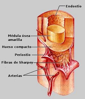
The Sharpey fibers They are a set of collagen extensions that form a strong, low mineralized network that firmly binds bones to muscles and ligaments. They are also found on the external surface of the bone, where it is responsible for attaching the bone to the periosteum..
These fibers have been the subject of study over the years since their function and their adaptation mechanism to the bone environment were not well understood. From experiments in rodents it has been possible to better study its structure, its function and its development.
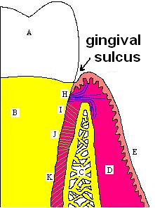
In teeth, Sharpey's fibers are the terminal branches of the periodontal ligament, which cuts through the dental cementum to join the tooth with the periosteum of the alveolar bone of the jaws..
Sharpey's fibers were long thought to be inert and did not have any change during the stages of bone resorption and renewal, however there is current evidence that they are capable of varying their size and diameter to accommodate bone metabolism.
Article index
Sharpey's fibers are filaments of collagen and other elements that support the skeletal system with the periosteum and the muscles and ligaments.
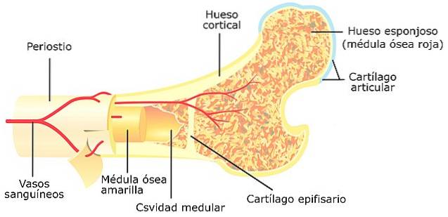
The bones have an external surface, which is covered by a fibrous sheet called periosteum. This membrane is rich in blood vessels and neurological endings; provides a good part of the external vascularization of the bone.
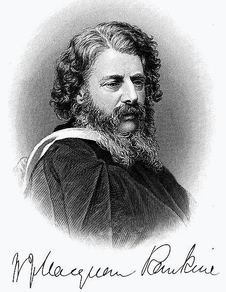
In the 1867 edition of the publication Elements of anatomy, Dr. William Sharpey described the existence of a complex fibro-elastic network of collagen, which pierced the bone and reached the periosteum, strongly joining these structures. These same fibers were present at the attachment of bones to muscles and ligaments.
By 1923, these branches of collagen were already known as Sharpey's fibers. That same year its presence was observed on the bony surface of the teeth.
In 1972, Dr. Cohn studied the internal composition of the tooth with an emphasis on the Sharpey fibers, describing their path from the dental cementum to the alveolar bone of the maxilla..
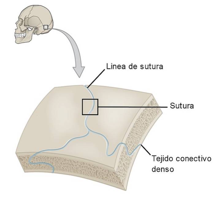
Sharpey's fibers are also present between the bones of the skull. Forming firm but elastic partings.
Most of the research known on Sharpey's fibers has focused on studying them from those that are part of the dento-alveolar organization.
Previously, it was thought that these perforating fibers were a suspension network formed only by collagen, however, this theory is ruled out since immunohistochemical studies have shown that their structure is much more complex..
In addition, the way in which this matrix maintained its fibrous consistency, escaping the calcification caused by bone mineral elements, was striking..
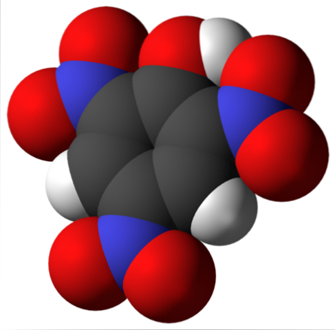
Sharpey fibers are currently known to be composed of type III and VI collagen, elastin, and glycoproteins tenascin Y fibronectin.
The association of type III collagen with type VI collagen provides great stability to the Sharpey fiber network, which explains its firmness during the bone remodeling stages..
Studies carried out on the fibers located in the teeth have been able to differentiate between two types of fibers depending on their thickness: thick and fine. The thick ones measure between 8-25 µm and the thin ones less than 8 µm.
Sharpey fibers are responsible for establishing strong bonds between the bone surface and the periosteum, muscles and ligaments.
However, it is known that in addition to this function, its complex protein structure plays a fundamental role in bone formation during the fetal stage, in increasing bone resistance in athletes and in bone repair in the event of trauma or injury. physiological damage.
At the time of bone formation, during gestation, the network of Sharpey fibers is formed around primitive bones..
Collagen fibers with elastin and glyuproteins tenascin and fibronectin, organize themselves by emitting signals for cell migration and differentiation of bone cells.
When there are problems in the structure of the Sharpey fibers, pathologies of bone formation such as fibrous dysplasia, in which the primitive bones do not finish calcifying properly.
In menopausal patients, there is a decrease in bone mineralization that results in calcium loss and osteoporosis..
As for the Sharpey fibers, their organization is affected by the hormonal decrease causing their decrease in some areas of the bone..
This situation makes these areas more susceptible to mineral loss and, as a consequence, to osteoporosis..
Likewise, it is believed that the progressive muscle atrophy seen in these types of patients is in part due to the decrease in the population of Sharpey fibers that hold bone to muscle..
The hormonal signals that are triggered when there is bone damage, and that activate the pathways for repair from the bone cells, also activate an adaptation mechanism in the Sharpey fibers.
Damage to the periosteum elongates the collagen in the fibers, causing them to begin to increase in diameter and size to prepare for the stage of new bone tissue formation..
Once bone remodeling is complete, the fibers return to their original size and arrangement.
The amount of Sharpey fibers has been observed to be increased up to 7% more in people who engage in physical activity, such as running, compared to those who are sedentary.
This increase has benefits in terms of bone resistance and the proper functioning of the joints..
As time passes, Sharpey fibers, like other elements, change their protein structure, substituting type III collagen for type I collagen..
The union of type I collagen with type VI collagen does not have the same resistance effect as the original alliance, so a process of wear begins that ends in the calcification of some of the fibers of the protein network.
These calcifications make the joints not as firm as they should be. In the case of teeth, there may be movement of the same and even fall due to not having a stable support form.

Yet No Comments