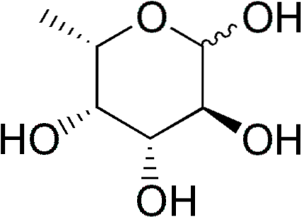
The fucose (abbreviated Fuc), or 6-L-deoxy-galactose, is a partially deoxygenated (deoxysugar) six-carbon monosaccharide whose empirical formula is C6H12OR5. Like other monosaccharides, it is a polyhydric sugar.
When a hydroxyl group is replaced by a hydrogen atom, a deoxysugar is derived. Although theoretically this replacement could affect any hydroxyl group of any monosaccharide, in nature there is little variety of deoxysugars.
Some deoxysugars are: 1) deoxyribose (2-deoxy-D-ribose), derived from D-ribose, which is part of DNA; 2) rhamnose (6-D-deoxymannose), derived from D-mannose; 3) fucose, derived from L-galactose. The latter is more common than D-fucose, derived from D-galactose.
Article index
Fucose is also known by the names 6-deoxy-galacto-hexose, fucopyranose, galactomethylose, and rodeose..
Although normally found in forming polysaccharides and glycoproteins, isolated as a monosaccharide it is sweeter than galactose. This is due to the fact that the replacement of a hydroxyl group by a hydrogen atom increases the hydrophobic character and, therefore, the sweetness of the molecule..
The hydroxyl groups of fucose can undergo the same reactions as other sugars, producing a wide variety of acetals, glycosides, ethers and esters..
A fucosylated biomolecule is one to which, by the action of a fucosyltransferase, fucose molecules have been attached through glycosidic bonds. When the hydrolysis of glycosidic bonds occurs by the action of a fucosidase, thus separating the fucose, the biomolecule is said to have been defucosylated.
As glucans are fucosylated, more complex glucans called fucans are generated, which may or may not be part of glycoproteins. Sulfated fucans are defined as those polysaccharides that contain sulfated L-fucose residues. They are typical of brown algae. Examples include ascophylane, sargasan, and pelvetan..
One of the best-studied fucans is fucoidan, obtained from the brown alga Fucus vesiculosus, which has been marketed (Sigma-Aldrich Chemical Company) for decades.
D-fucose is present in antibiotic substances produced by microbes, and in plant glycosides, such as convolvulin, chartreusin, ledienoside and keirotoxin.
L-fucose is a constituent of polysaccharides from algae, plum leaves, flax, soy and canola seeds, gum tragacanth, potato cell walls, cassava tubers, kiwi fruit, the bark of the ceiba and the mucigel of the corn caliptra, as well as other plants.
L-fucose is also present in sea urchin eggs and in the gelatin that protects frog eggs.
In mammals, fucans with L-fucose form the ligands that act on selectin-mediated leukocyte-endothelial adhesion, and participate in numerous ontogenetic events.
L-fucose is abundant in the fucosphingolipids of the gastrointestinal epithelium and bone marrow, and appears in small proportions in cartilage and keratinous structures..
In humans, fucans with L-fucose are part of glycoproteins in saliva and gastric juices. They are also part of the antigens that define ABO blood groups. They are present in various oligosaccharides in breast milk.
Fucosyltransferases use GDP-fucose, a nucleotide-activated form of fucose, as a fucose donor in the construction of fucosylated oligosaccharides.
GDP-fucose is derived from GDP-mannose by the successive action of two enzymes: GDP-mannose 4,6-dehydratase and GDP-4-keto-6-deoximanose 3,5-epimerase-4-reductase.
Using a NADP + cofactor, the first enzyme catalyzes the dehydration of GDP-mannose. Reduction of position 6 and oxidation of position 4 produces GDP-6-deoxy-4-keto-mannose (during the reaction, the hybrid is transferred from position 4 to 6 of the sugar).
The second enzyme, which is NADPH dependent, catalyzes the epimerization of positions 3 and 5, and the reduction of the 4-keto group, of GDP-6-deoxy-4-keto-mannose..
Bacteria can grow using fucose as the sole source of carbon and energy by means of a fucose-inducible operon that encodes catabolic enzymes for this sugar..
The above process involves: 1) entry of free fucose through the cell wall mediated by a permease; 2) isomerization of fucose (an aldose) to form fuculose (a ketosis); 3) phosphorylation of fuculose to form fuculose-1-phosphate; 4) an aldolase reaction to form lactaldehyde and dihydroxyacetone phosphate from fuculose-1-phosphate.
Symptoms of many types of cancerous tumor include the presence of glucan-bound proteins that are distinguished by having an altered oligosaccharide composition. The presence of these abnormal glucans, among which fucans stand out, is linked to the malignancy and metastatic potential of these tumors.
In breast cancer, tumor cells incorporate fucose into glycoproteins and glycolipids. Fucose contributes to the progression of this cancer, favoring the activation of cancer stem cells, hematogenic metastasis and the invasion of tumors through extracellular matrices..
In lung carcinoma and hepatocarcinogenesis, increased fucose expression is associated with a high metastatic potential and a low probability of survival.
In contrast, some sulfated fucans are promising substances in the treatment of cancer, as has been determined by numerous in vitro studies with cancer cell lines, including those that cause breast, lung, prostate, gastric, colon and rectal cancer..
Increased expression of fucans in serum immunoglobulins has been associated with juvenile and adult rheumatoid arthritis.
Leukocyte adhesion deficiency II is a rare congenital disease due to mutations that alter the activity of a FDP-fucose transporter located in the Golgi apparatus.
Patients suffer from mental and psychomotor retardation, and suffer from recurrent bacterial infections. This disease responds favorably to oral doses of fucose.
Sulfated fucans obtained from brown algae are important reservoirs of compounds with therapeutic potential.
They have anti-inflammatory and antioxidant properties, inhibiting the migration of lymphocytes at sites of infection and favoring the release of cytokines. Increase the immune response by activating lymphocytes and macrophages.
They have anticoagulant properties. Orally shown to inhibit platelet aggregation in human patients.
They have antibiotic and antiparasitic potential and inhibit the growth of stomach pathogenic bacteria Helicobacter pylori. Kill parasites Plasmodium spp. (causative agent of malaria) and Leishmania donovani (causative agent of American viscerotropic leishmaniasis).
Finally, they have powerful antiviral properties, inhibiting the entry into the cell of several viruses of great importance to human health, including Arenavirus, Cytomegalovirus, Hantavirus, Hepadnavirus, HIV, herpes simplex virus, and influenza virus.


Yet No Comments