
The protein glycosylation It is a post-translational modification that consists of the addition of linear or branched oligosaccharide chains to a protein. The resulting glycoproteins are generally surface proteins and secretory pathway proteins..
Glycosylation is one of the most common peptide modifications among eukaryotic organisms, but it has been shown to occur in some species of archaea and bacteria as well..
In eukaryotes, this mechanism occurs between the endoplasmic reticulum (ER) and the Golgi complex, with the intervention of different enzymes involved both in regulatory processes and in the formation of protein + oligosaccharide covalent bonds..
Article index
Depending on the binding site of the oligosaccharide to the protein, glycosylation can be classified into 4 types:
It is the most common of all and occurs when oligosaccharides bind to the nitrogen of the amide group of asparagine residues in the Asn-X-Ser / Thr motif, where X can be any amino acid except proline.
When carbohydrates bind to the hydroxyl group of serine, threonine, hydroxylysine, or tyrosine. It is a less common modification, and examples are proteins such as collagen, glycophorin, and mucins..
It consists of the addition of a mannose residue that binds to the protein through a C-C bond with the C2 of the indole group in tryptophan residues.
A polysaccharide acts as a bridge to attach a protein to a glycosylphosphatidylinositol (GPI) anchor on the membrane.
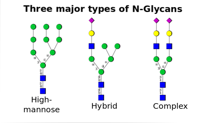
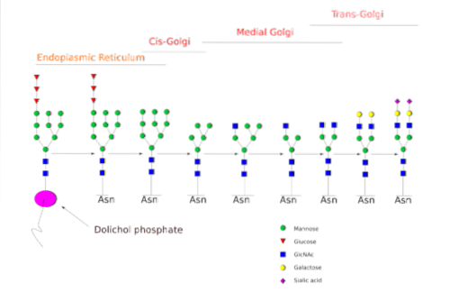
The N-glycosylation is the one that has been studied in more detail. In mammalian cells, the process begins in the rough ER, where a preformed polysaccharide binds to proteins as they emerge from ribosomes..
Said precursor polysaccharide is composed of 14 sugar residues, namely: 3 glucose (Glc), 9 mannose (Man) and 2 N-acetyl glucosamine (GlcNAc) residues.
This precursor is common in plants, animals, and single-celled eukaryotic organisms. It is bound to the membrane thanks to a bond with a dolichol molecule, an isoprenoid lipid embedded in the ER membrane..
After its synthesis, the oligosaccharide is transferred by the oligosacryltransferase enzyme complex to an asparagine residue included in the tri-peptide sequence Asn-X-Ser / Thr of a protein while it is being translated.
The three Glc residues at the end of the oligosaccharide serve as a signal for correct oligosaccharide synthesis, and are cleaved along with one of the Man residues before the protein is carried into the Golgi apparatus for further processing..
Once in the Golgi apparatus, the oligosaccharide portions attached to the glycoproteins can be modified by the addition of galactose, sialic acid, fucose, and many other residues, yielding chains of much greater variety and complexity..
The enzymatic machinery that is needed to carry out the glycosylation processes includes numerous glycosyltransferases for the addition of sugars, glycosidases for their removal, and different nucleotide sugar transporters for the contribution of the residues used as substrates..
Bacteria do not have intracellular membrane systems, so the formation of the initial oligosaccharide (of only 7 residues) occurs on the cytosolic side of the plasma membrane..
Said precursor is synthesized on a lipid that is then translocated by an ATP-dependent flipase into the periplasmic space, where glycosylation occurs..
Another important difference between eukaryotic and prokaryotic glycosylation is that the enzyme oligosaccharide transferase (oligosacaryltransferase) from bacteria can transfer sugar residues to free portions of already folded proteins, not as they are translated by ribosomes..
Furthermore, the peptide motif recognized by this enzyme is not the same eukaryotic tri-peptide sequence..
The N-oligosaccharides attached to glycoproteins serve various purposes. For example, some proteins require this post-translational modification to achieve the proper folding of their structure..
To others it provides stability, either by avoiding proteolytic degradation or because this portion is necessary for them to fulfill their biological function..
Since oligosaccharides have a strong hydrophilic character, their covalent addition to a protein necessarily modifies its polarity and solubility, which may have relevance from a functional point of view..
Once attached to membrane proteins, oligosaccharides are valuable information carriers. They participate in the processes of cell signaling, communication, recognition, migration and adhesion.
They have an important role in blood clotting, healing and immune response, as well as in the processing of protein quality control, which is glycan-dependent and indispensable for the cell..
At least 18 genetic diseases have been linked to protein glycosylation in humans, some of which involve poor physical and mental development, while others can be fatal.
There are a growing number of discoveries related to glycosylation diseases, especially in pediatric patients. Many of these disorders are congenital and have to do with defects associated with the initial stages of oligosaccharide formation or with the regulation of the enzymes that participate in these processes..
Since a large part of the glycosylated proteins make up the glycocalyx, there is a growing interest in verifying that mutations or alterations in the glycosylation processes may be related to the change in the microenvironment of tumor cells and thus promote the progression of tumors and development of metastases in cancer patients.


Yet No Comments