
GLUT2 It is a low-affinity glucose transporter that is expressed in the membranes of pancreatic, liver, kidney, and intestinal cells as well as in astrocytes and tanicytes. In addition to mediating glucose transport, it is also involved in the transport of fructose, galactose, and glucosamine; so more than a glucose transporter it is a hexose transporter.
Its low affinity for glucose allows it to act as a sensing protein for blood glucose levels. Therefore, it participates in the regulatory control of many physiological events that respond to fluctuations in the concentration of glucose in the blood..
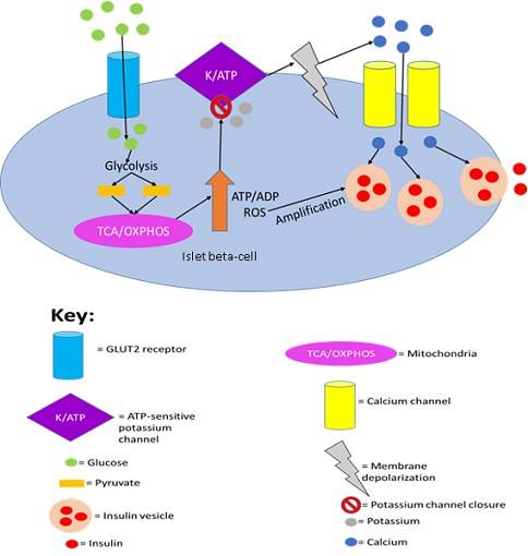
Among the many processes that it regulates, the following stand out: 1) the release of insulin by pancreatic cells stimulated by high concentrations of glucose; 2) glucagon secretion by hepatocytes for glucose production in hypoglycemia.
Article index
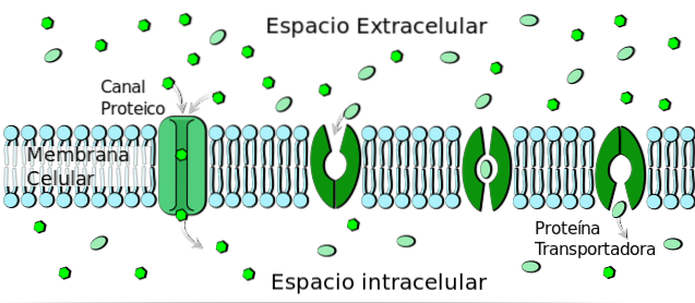
Approximately 75% of glucose that enters the cell to fuel metabolic pathways for energy production does so through a passive transport mechanism facilitated by integral membrane proteins called transporters..
This transport mechanism is widely known as facilitated diffusion. It does not require a contribution of energy to be carried out and it is given in favor of a concentration gradient. That is, from an area of high concentration to one of low concentration.
At least 14 isoforms of glucose facilitated diffusion transporters, including GLUT2, have been identified to date. All of them belong to the main superfamily of facilitators (MSF) and, by consensus, called GLUTs (for the acronym in English of “Glucose Transporters”).
The different GLUTs that have been characterized to date are encoded by SLC2A genes and exhibit marked differences in amino acid sequence, preference for transporting substrates, and cellular and tissue distribution..
GLUT2 mobilizes glucose through a transport mechanism in a single direction (uniport). This function is also performed by GLUT1, the most abundant glucose transporter in practically all mammalian cells..
However, unlike this, it has an extremely low affinity for glucose, which means that it is only capable of transporting it when the concentrations of this sugar tend to reach very high values in the extracellular environment..
Despite having a low affinity for glucose, it has a high transport capacity, which implies that it can transport large amounts of this hexose at high speed. These two characteristics appear to be related to the role of this transporter in responding to subtle changes in glucose concentration..
Molecular characterization studies of this transporter have shown that it does not have unique specificity for glucose. On the contrary, it is able to mediate the passive transport of fructose, galactose, mannose and glucosamine. Presenting low affinity for the first three and high affinity for glucosamine.
Since all these molecules are sugars with six carbon atoms, it can be considered as a hexose transporter rather than a glucose transporter..
GLUT2 has a peptide sequence 55% identical to that of the high affinity transporter for glucose GLUT1.
However, despite this low percentage of similarity between the sequences of both transporters, studies carried out by X-ray crystallography have shown that they present a similar structure..
This structure corresponds to that of an α-helix multipass transmembrane protein. That is, it crosses the membrane multiple times through transmembrane segments that present α-helix configuration..
As in all the members of the main super family of facilitators (MSF), to which it belongs, 12 helical segments cross the membrane. Six of these rearrange themselves spatially to form a hydrophilic pore through which sugars are mobilized..
It should be noted that the hexose binding site is defined by the orientation and pseudopsymmetry presented by the carboxyl and amino terminal ends of the protein. Both exposed to the same side of the membrane generate a cavity in which the arrangement of the six sugar atoms are recognized, facilitating their union..
A change in the structure of the transporter is related to the mechanism used by it to transport sugars from one side of the membrane to the other. This structural deformation makes it possible to mobilize the binding site towards the cytoplasmic side, where the release of the molecule that has been transported rapidly occurs.
In addition to mediating the sequestration of glucose, mannose, galactose and glucosamine within the cell, numerous physiological functions have been attributed to the expression of this transporter in various cell types.
Many of these functions have been determined using gene suppression techniques. The latter consist of preventing the expression of the gene whose function is to be studied in the cells of a specific tissue or of a complete organism..
In this sense, blocking the expression of GLUT2 in mice has revealed that this protein constitutes the main means of glucose transport in both kidney and liver cells. In addition, the transport of galactose and fructose is not related to the generation of glucose from these sugars via gluconeogenesis..
Additionally, it has been shown that it exerts a regulatory role in various physiological functions, since its low affinity for glucose allows it to detect when the concentrations of this sugar are high..
Since it plays a critical role in the generation of energy by all cells, especially nerve cells, its concentration in the blood must be kept close to a value of 5mmol / l. Variations in this concentration are always being monitored by regulatory proteins through "glucose detection" mechanisms..
These mechanisms consist of molecular strategies that allow a rapid response to sudden variations in glucose concentration. In this sense, the expression of GLUT2 in the membrane of cells whose functions are activated by hyperglycemia confers on it a regulatory role..
In fact, it has been shown that insulin secretion by pancreatic cells is triggered by the detection of glucose by GLUT2..
Additionally, it mediates the autonomic nervous control of feeding, thermoregulation and the functioning of pancreatic cells stimulated by glucose detection..
When GLUT2 levels decrease in nerve cells they generate a positive signal to trigger glucagon secretion. Remembering that glucagon is a hormone that promotes glucose production by the liver from glycogen stores.

Yet No Comments