The granulopoiesis It is the process of renewal of the granulocytic cells that circulate in the blood and that are part of the body's defense system. Granulopoiesis occurs in the bone marrow. This process includes the formation and maturation of granulocytic cells such as the segmented neutrophils, eosinophils, and basophils..
Blood cells arise from a pluripotential stem cell that differentiates into various cell lines; and these in turn differentiate into slightly more differentiated cell lines, until they reach circulating mature cells.
During the granulopoiesis process, cells undergo a series of changes as they differentiate into more mature cells..
The most notable changes are:
- Decreased size of cells.
- Decreased nucleus-cytoplasm ratio (smaller nucleus and larger cytoplasm).
- Core condensation and fragmentation.
- Invisibility of the nucleoli.
- Appearance of primary and later secondary granules in the cytoplasm.
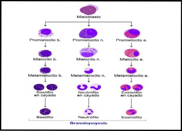
The primary granules are azurophilic and later become acidophilic, neutrophilic or basophilic, depending on the cell line to which it belongs. Granulocytic cells go through various stages of differentiation: myeloblasts, promyeloblasts, myelocytes, metamyelocytes, arch (banded nucleus), and mature granulocytes.
This process is regulated by stimulating and inhibiting substances produced by the cells of the immune system..
Article index
The process of forming all blood cells is called hematopoiesis. Therefore, granulopoiesis is part of hematopoiesis.
Granulopoiesis represents the formation and maturation of a specific group of cells that make up 60% of blood cells.
The complete kinetics of granulocytes include the formation, maturation, circulation and redistribution in organs and tissues..
This means that granulopoiesis is not a static process, since during the formation and maturity process the cells migrate towards various compartments inside and outside the bone marrow..
The compartments described are 4 and are mentioned below:
- Formation and maturation.
- Backup.
- Circulating.
- Of marginalization
These compartments have been extensively studied, based on the kinetics of the segmented neutrophil as it is the most abundant granulocyte in the blood..
The first two compartments develop in the bone marrow. The granulocyte formation and maturation process lasts approximately 11 days, of which the granulocytes spend 7 days in the formation and maturation compartment and then go to the reserve compartment, where they remain for 4 days..
When the segmented neutrophils leave the reserve compartment and enter the circulation, a percentage of them will travel freely in the blood. However, others will adhere to the walls of the capillaries and post-capillary venules or will be retained in capillaries close to the great veins. This is what is known as a compartment of marginalization.
Granulocytes have a half-life of 6 to 8 hours. Therefore, to maintain homeostasis for the number of granulocytes in the blood, the bone marrow must produce billions of granulocytes per day..
In this sense, the granulocytes that are destroyed in organs and tissues are quickly replaced thanks to the marginalization and reserve compartment..
There are physiological causes that can increase the number of segmented neutrophils, without there being an increase in production. This happens, for example, during physical exercise. In addition, in case of bacterial infections, the production of granulocytes increases, while the stay of these cells in the reserve compartment decreases..
In pathological processes such as leukemias, there is a lack of control in the formation, maturation and distribution of cells, which is why an exorbitant number of immature cells will be observed in circulation..
The count and differentiation of leukocytes is a very important parameter in complete hematology. The leukocyte count provides guidance on the immunological status of the patient, in addition to providing data that help to show infectious processes or malignant diseases.
In the special case of granulocytes, these provide extremely important data, since bacterial infections are characterized by leukocytosis and neutrophilia. That is, an increase in the total number of leukocytes and an increase in the number of segmented neutrophils, respectively..
While in viral infections they present with leukopenia (decrease in the total number of leukocytes) and with neutropenia (decrease in the number of segmented neutrophils).
Likewise, segmented eosinophils tend to increase in allergic and parasitic processes..
In the blood smear, mature granulocytes can be observed and quantified, that is, segmented neutrophils, eosinophils and basophils..
The characteristics of these cells are as follows.
It measures between 9 and 12 µm. It is the most abundant granulocytic cell in the blood, and normally reaches a percentage of 60 to 70% in the blood circulation (normal value). Its cytoplasm is acidophilic and contains abundant neutrophilic granules.
The nucleus usually takes various forms, and as its name indicates it is segmented into 2 to 5 lobes. The more lobes it has, the older the cell.
Therefore, some bioanalysts and hematologists, based on Arneth's scheme, report “formula deviated to the left” when neutrophils with few lobulations predominate, and “formula deviated to the right” when they present a greater number of lobulations..
This cell is easily recognizable for its peculiar characteristics. It is characterized by having a nucleus with two clearly visible lobulations and by presenting abundant and thick acidophilic granulations in its cytoplasm, without covering the nucleus..
Segmented eosinophils are found in low concentrations in peripheral blood, their normal value being between 1 to 3%. This increases in allergic processes and in some parasitosis.
These cells are the ones with the fewest numbers: the normal value in the blood ranges from 0 to 1%. They are characterized by having a polymorphic nucleus and a cytoplasm full of thick basophilic granulations that are superimposed on the nucleus, preventing its visualization..
The process of formation and maturation of granulocytes goes through various stages or phases.
From the multipotential hematopoietic stem cell (hemocytoblast) the myeloid precursor cell is generated, and this in turn gives rise to the granulocytic / monocytic progenitor cell, which later gives rise to the myeloblast.
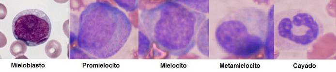
This cell measures 14 to 20 µm and is characterized by an oval nucleus that covers almost the entire cell. Therefore, its cytoplasm is scarce. Its chromatin is lax, being able to appreciate 1 to 3 nucleoli.
The myeloblast has a basophilic cytoplasm, and no granulations are observed. This cell divides to give rise to two promyelocytes..
The promyelocyte is the cell that continues after the myeloblast stage. The nucleus presents a slightly denser chromatin, however it is still possible to observe the nucleoli.
Despite the fact that in the maturation process the rule is that the size of the cell decreases, in this case the promyelocyte is the largest cell. Measures between 16-25 µm.
The nucleus is smaller, showing more cytoplasm. This is still basophilic and presents azurophilic granules (primary granulations).
This cell measures 12 to 18 µm and has a more advanced degree of maturation than the promyelocyte. The nucleus can be seen oval or with a pronounced cleft, and the shape can even become kidney-shaped.
The chromatin becomes denser and the nucleoli can no longer be seen. The cytoplasm becomes slightly acidophilic, and secondary granules appear that reveal the type of granulocyte that is maturing (eosinophils, neutrophils or basophils).
At this stage the nucleus is eccentric and is characterized by a deeper cleft. A more condensed chromatin is observed compared to the previous stage.
In this degree of maturation, specific granules are abundant according to the type of granulocyte that is developing, while the primary granules still present are no longer visible..
At this stage the cell loses the property of dividing. Under certain conditions (severe bacterial infections) they could be seen circulating in the blood in low amounts, without representing a serious myeloid disorder..
However, if it is found in high amounts, it indicates a pathological process called myeloid leukemia..
This stage is only observed in the case of the maturation of the segmented neutrophils. It is also known as a juvenile neutrophil.
It can be seen circulating in the blood under specific conditions, such as in bacterial infectious processes in which there is a significant increase in the number of circulating leukocytes at the expense of segmented neutrophils (marked neutrophilia).
This cell is characterized by presenting a band-shaped nucleus that simulates the letter "C" or a horse's shoe. On the other hand, abundant neutrophil granules and few azurophils are found in the cytoplasm.
These comprise the 3 types of granulocytes found in the peripheral blood. These are: segmented neutrophils, segmented eosinophils, and segmented basophils. Its characteristics have already been described in the hematology section.
Granulopoiesis is regulated by certain substances that are synthesized by cells of the immune system, such as lymphocytes, macrophages, and the granulocytic cells themselves..
Some have stimulating and other inhibitory functions. Therefore, these substances maintain the balance of cell clones and the proper functioning of the immune response..
Although it is still unknown what stimuli the pluripotential stem cell receives to divide and differentiate into precursor cells of the lymphoid and myeloid line, it is believed that interleukin 3 (IL3-) produced by CD4 lymphocytes could act in this sense, in addition to other signals they receive from the medullary microenvironment.
Likewise, the granulo-monocytic colony stimulating factor (GM-CSF) is found, which stimulates the precursor cell of the myeloid series to originate the granulocytic / monocytic progenitor cell.
Granulocytic colony stimulating factor (G-CSF) is also found, which stimulates the maturation of the precursors of segmented neutrophils, eosinophils and basophils..
Interleukin (IL 5) intervenes for the differentiation of the segmented eosinophils, while eotaxin 1 acts on the migration and recruitment of eosinophils..
A substance released by the granules of segmented neutrophils called lactoferrin participates in the regulation of the expression of genes involved in various functions of cell activation and cytokine production..
In this sense, lactoferrin acts by activating macrophages, which can release prostaglandin-E(PGE). This substance, together with chalones and acidic isoferritins, inhibit the proliferation and maturation of segmented neutrophils..
On the other hand, lactoferrin stimulates the activation of CD4 lymphocytes and the release of anti-inflammatory Interleukin (IL-10).


Yet No Comments