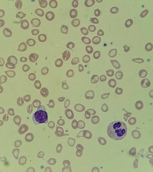
The hypochromia It is the decrease in color or paleness of erythrocytes or red blood cells. The cause of this reduction in coloration is the decrease in the concentration of hemoglobin within the red blood cells, which decreases the transport of oxygen in the blood and produces a pathophysiological condition called “anemia”.
Alterations in red blood cell function include alteration in the number of circulating erythrocytes or alterations in their components, including hypochromia.
Anemia strictly refers to a decrease in the number or volume of circulating red blood cells, or a decrease in the quality or quantity of hemoglobin contained within these cells..
Anemias can be the result of problems in the formation of red blood cells and / or hemoglobin, acute or chronic blood loss, increased destruction of red blood cells, or a combination of these factors..
Anemias are classified according to their etiology or according to their morphology. The morphological classification, which is the one related to hypochromia, has to do with globular size and hemoglobin content.
Globular size is identified by the suffix “citic” and the hemoglobin content is identified by the suffix “cromic”. This is how hypochromic, normochromic and hyperchromic anemias are described; microcytic, normocytic and macrocytic. Hypochromia is generally related to hypochromic microcytic anemias.
Article index
It has already been explained that anemia is related to a decrease in hemoglobin or the number of red blood cells. A decrease in the number of these cells causes, in turn, a decrease in circulating hemoglobin and a reduction in the hemoglobin content in each red blood cell has the same effect..
Hemoglobin is a protein contained within the erythrocyte and whose function is to serve as an oxygen transport molecule in the blood.
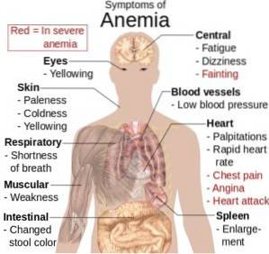
The clinical manifestations of anemia are due to a decrease in the ability of the circulating blood to transport oxygen to the tissues and the possible production of tissue hypoxia (decreased oxygen supply to the tissues).
The symptoms of hypochromic anemia are very varied and depend on the severity of the anemia and the ability of the body to compensate for this reduced capacity..
If the anemia develops gradually and the reduction in red blood cells or hemoglobin is moderate, the compensatory mechanisms can be so efficient that there are no symptoms at rest, but these appear during periods of physical exercise.
As the loss of erythrocytes or hemoglobin continues, the symptoms become evident and the compensatory alterations of some organs and systems are frank. The systems involved in compensation are the cardiovascular system, the respiratory system, and the hematological or hematopoietic system..
If compensatory mechanisms fail, dyspnea (shortness of breath), tachycardia, palpitations, throbbing headache, dizziness, and fatigue quickly appear, even at rest. Decreased oxygen supply to skeletal and muscle tissue can lead to pain, claudication, and angina.
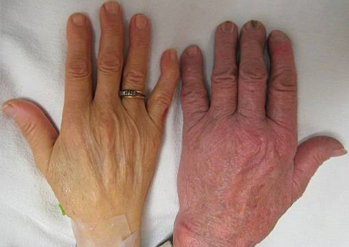
When hemoglobin levels are between 7 and 8%, intense paleness appears in the palms of the hands and feet, in the skin and mucous membranes (especially in the ocular conjunctiva), as well as in the earlobes. Nails become shiny, thin and brittle, with a spoon-shaped concavity (koilonicia) due to capillary deficit.
The tongue becomes red, painful, edematous, and shows papillary atrophy. The intensity of pain (glossodynia) is related to the degree of iron deficiency as the cause of anemia.
Hypochromic microcytic anemias can occur from several causes, including:
- Alterations in iron metabolism.
- Failures in the synthesis of porphyrins and group "heme".
- Globin synthesis failures.
Among these alterations, some specific causes such as iron deficiency anemia, sideroblastic anemia and thalassemias can be named..
Worldwide, iron deficiency anemia (hypoferremia) is the most common. There are some predisposing conditions, such as being mothers and nursing children who live in a state of chronic poverty.
In developed countries it is related to pregnancy and chronic blood loss due to duodenal or gastric ulcers, as well as neoplasms.
Physiopathologically, three stages are described in iron deficiency anemia. The first is where iron stores are depleted, but adequate hemoglobin synthesis is achieved. In the second stage, the supply of iron to the bone marrow decreases and hematopoiesis is altered.
In the third stage, finally, there is a decrease in the synthesis of hemoglobin and hypochromia appears.
It is a heterogeneous group of disorders characterized by anemias that vary in severity and are due to an inefficient absorption of iron, causing, consequently, the synthesis of a dysfunctional hemoglobin.
The presence of ringed sideroblasts in the bone marrow gives the diagnosis of sideroblastic anemia. Ringed cideroblasts are erythroblasts that contain iron granules that do not participate in the synthesis of hemoglobin and form a collar around the nucleus..
Several inherited and acquired causes are described. In the case of acquired ones, some are reversible, such as those related to alcoholism, with the reaction to certain drugs, with a copper deficiency and with hypothermia. Other acquired conditions are idiopathic and others are related to myeloproliferative processes (uncontrolled proliferation of hematopoietic cells).
Hereditary forms only occur in men, as they are related to recessive transmission on the X sex chromosome.
With the name of "thalassemia" it is grouped to a very heterogeneous set of congenital alterations whose common characteristic is that of having a defect in the synthesis of one or more globin chains. They are due to mutations in the genes that code for globin chains, which decrease their synthesis.
Thalassemias can affect the alpha chain or the beta chain of globin, which is why they are called "alpha" or "beta" thalassemias, respectively..
When the synthesis of one chain decreases, the other accumulates; thus in alpha thalassemias beta chains accumulate and in beta thalassemias alpha chains accumulate. They are related to severe anemia, are quite frequent and have an autosomal dominant inheritance pattern..
After making the diagnosis, when the cause is iron deficiency, the injuries that cause acute or chronic blood loss must be corrected. Iron supplements are started and hemoglobin levels quickly recover (1 to 2g / dl in the first weeks). This confirms the diagnosis of iron deficiency..
The most common form of administration of iron is as ferrous sulfate at a rate of 150 to 200mg / day and for a period of 1 to 2 months, being able to last up to three months.
Approximately one third of the cases of people with inherited sideroblastic anemia usually respond to pyridoxine treatment at a rate of 50 to 200 mg / day, although with variable responses. For those who do not respond to this treatment, transfusions are needed to ensure growth and development..
In thalassemias, treatment usually consists of a regimen of transfusions as needed. Splenectomy (removal of the spleen) is sometimes necessary.
Yet No Comments