
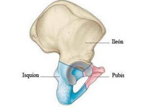
The hip bone It is a paired bone articulated posteriorly with the sacral bone of the vertebral column, and anteriorly with its contralateral counterpart through the symphysis pubis. This bone forms the pelvic girdle. It is the result of the union of three primitive bone pieces: the ilium, the ischium and the pubis; these converge in the acetabular fossa.
At the time of birth, this convergence is evidenced in the acetabulum in the form of three cartilaginous sheets arranged in a “Y” shape, which disappears in adults due to the ossification of the sheets. The hip bone is located between the lower abdomen and the upper part of the lower limbs.
The innominate bone is a deep bone that becomes shallower at four points: on both sides of the iliac crest, on both sides of the anterior superior iliac spines, on the underside of the pubic spine, and on the back of the ischial tuberosity..
Article index
It is the main constituent of the pelvis, together with the sacrum and coccyx, with which it articulates posteriorly.
One of the characteristics of the innominate bone is its constitution as a true flat bone, with two sheets of compact bone covering the cancellous bone..
Some parts are thinner than others. The thickest correspond to areas of firm muscle attachments, such as the iliac crest, the ischial tuberosity and the pubis..
Two faces, four edges and four angles are described in the innominate bone.
The most characteristic structure that can be found on the lateral aspect of the innominate bone is a wide, round and deep joint cavity called the acetabulum, which is circumscribed by the acetabular border..
This joint cavity has two parts: a non-articular square, called the acetabular fossa; and a joint that surrounds the crescent-shaped fossa, called the semilunar facet.
Two lines can be seen above the acetabulum: the anterior gluteal line and the posterior gluteal line. These divide the gluteal aspect of the bone into three regions:
- Posterior region, for gluteus maximus insertion.
- Middle region, for the insertion of the gluteus medius.
- Anterior region, for the insertion of the gluteus minimus.
This medial face is divided into two regions by the arcuate line, which is also called innominate and which is directed from top to bottom and from back to front..
- A superolateral region called the iliac fossa, which is smooth and serves as an insertion point for the iliac muscle.
- An inferomedial region where the iliac tuberosity can be seen, as well as various depressions and elevations intended for muscular and ligamentous insertion.
This border has a vertical portion oriented downwards that then abruptly changes position, becoming horizontal towards the medial. The characteristic elements of this edge are the following:
It results from the confluence of the iliac crest with the anterior border, into which the inguinal ligament, the tensor fascia lata muscle and the broad muscles of the abdomen are inserted..
Also called Freyggang notch. Immediately below the superior anterior iliac spine, it gives way to the lateral femoral cutaneous nerve.
In this projection the tendon of the rectus femoris is inserted.
The muscle passes through this depression on its way to insertion into the femur.
It is rounded below the depression of the iliopsoas muscle, into which the iliopectineal arch inserts.
It is the continuation of the arcuate line. On the triangular pectineal surface the pectineus muscle is inserted.
There is a protruding tubercle, the pubic spine, where the inguinal ligament is inserted.
It is the medial to the pubic spine, in which the rectus abdominis muscle and the pyramidal muscle are inserted.
It has an almost vertical direction and the following characteristic elements are clearly differentiated:
There the multifidus muscle is inserted and the posterior sacroiliac ligaments are attached..
It has no special clinical and topographic connotation.
A great variety of vessels and nerves pass through this, as well as the piriformis muscle, superior gluteal vessels and nerves, sciatic and inferior gluteal nerves, internal pudendal vessels and nerves, among others..
It is arranged in the shape of a triangular eminence. The sacrospinous ligament is inserted at its vertex, the superior gastrocnemius muscle is inserted on its lateral aspect and the posterior fascicles of the levator ani muscle are attached to its medial aspect..
The internal obturator muscle and the internal pudendal vessels and nerves pass through there..
Corresponds to the lower angle of the bone.
It is linked to the iliac crest, which, seen from above, is shaped like a S italic: thick in front and back, and thin medially..
It has two sub-borders or lips, separated by a line in which the external oblique, internal oblique and transverse muscles of the abdomen are inserted..
The iliac tubercle, where the gluteus medius muscle attaches, is located behind the anterior superior iliac spine on the outer lip of the iliac crest.
It corresponds to the border that goes from the angle of the pubis - with the articular facet towards the contralateral pubic bone called the surface of the symphysis - to the body of the ischium.
The lower border of the innominate bone has numerous ridges that serve as an insertion for the corpora cavernosa of the penis or clitoris, as well as for various muscles such as the gracilis, the adductor magnus and the fascia of the perineum..
Corresponds to the anterior superior iliac spine.
Corresponds to the posterior superior iliac spine.
It is represented by the symphysis pubis.
It is represented by the ischial tuberosity, one of the most robust areas of the bone.
Its main function is to articulate the axial skeleton with the lower limbs, connecting the spine with the femur through the shoulder girdle..
It is one of the bones that receives the most muscle insertions, and is largely responsible for the transfer of mechanical forces from the body to the lower limbs..
Between the articular facet and the upper face of the acetabulum, a column of thick spongy tissue is visualized, which transmits resistance to the weight of the body in the orthostatic position..
By forming the bony pelvis, the articulated hip bone provides structural support for the abdominal and pelvic viscera, as well as for the pregnant uterus. At the same time, it helps protect the pelvic structures from trauma.


Yet No Comments