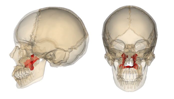
The palatine bone It is the name given to the bone structure that is above the palate and gives it its shape. Etymologically its name derives from the suffix "ino", which means "proper of"; and the word palatum, which means palate. In conjunction with other bone structures, this bone shapes the face in the human body..
Under normal conditions it is symmetrical and bilateral. The importance of anatomical knowledge of this structure is that its agenesis or alteration can generate serious aesthetic alterations with important psychological repercussions. In addition, it is the anatomical seat of many important vascular and muscular structures for man..
Article index
The palatine bone is a solid bone structure that is closely related to the upper jaw and plays a role in shaping the oral cavity..
Two major anatomical landmarks, the palatal lamina, a perpendicular lamina and a horizontal lamina are described in this.
It has four edges and two faces. It is quadrilateral in shape and constitutes the posterior part of the bony palate. In this sheet are the following parts:
Its posteromedial angle joins the same angle of the same border of the contralateral bone and forms the posterior nasal spine..
Joins the posterior border of the palatal process of the maxilla.
Inserts the vomer bone through the nasal crest at the top.
Follow the perpendicular sheet.
It is part of the floor of the nasal cavity.
Helps to form the vault of the bony palate.
Like the horizontal sheet, in its constitution it has two faces and four edges.
In turn, it has three zones: an anterior one, which contributes to the formation of the greater palatine groove; a posterior one, where the pterygoid process articulates; and an intermediate one, which forms the medial wall of the pterygopalatine fossa.
It has two ridges: one called the medial crest, which articulates with the middle nasal turbinate; and another called turbinal crest or crest of the shell.
It is superimposed on the process of the maxilla
Provides insertion to the soft palate. Articulates with the pterygoid process.
It has two processes, in the middle of which is the sphenopalatine notch.
In its anterior part the minor palatine canals are formed.
It articulates with 6 bones in total. These include the inferior turbinate, the vomer, the upper jaw, the sphenoid, the ethmoid and the contralateral palatine..
The two sheets that make up the palatine bone provide attachment to the following muscles:
Muscle whose main action is the elevation of the jaw.
Muscle whose main function is the protrusion of the jaw.
Muscle related to physiological swallowing.
In charge of maintaining the tension of the soft palate.
The soft palate descends.
Traction of the soft palate to one side.
Among the functions of this bone we can describe the following:
- Contribution in the formation of the nostrils.
- Act as a vocal sounding board when speaking.
- Provide symmetry to the face.
- Contribute to the formation of the palatal vault in the oral cavity.
- It is part of the constitution of the orbit and the pterygopalatine fossa.
Palatine bone pathologies are quite frequent. The most prominent are the following:
Embryologically, under normal conditions the lateral palatal fissures should fuse with the medial palatal fissures. If this does not occur, it gives rise to a clinical entity known as a cleft palate, where there is an opening in the palate.
These fissures can be incomplete when they only cover the soft palate, or complete when they cover the hard and soft palate. In this disease there is a direct communication between the nose and the mouth.
This disease presents important clinical manifestations that can severely affect the lives of the individuals who suffer from it. Some of its consequences are the following:
- Missing or delayed teething.
- Language development problems due to alteration of the speech apparatus.
- Feeding problems due to impaired chewing apparatus.
- Recurrent infections of the ear and nose, which is a notable problem since in the course of these diseases other more aggressive and potentially lethal clinical pictures could develop, such as meningitis.
The resolution of this pathology is purely surgical and must be carried out early.
Also called palatal, it is an abnormal bony growth on the surface of the palate, usually in the midline. They are generally no larger than 2 cm.
Its etiology is unknown, but there are hypotheses that argue that it is due to an autosomal dominant defect. However, it has been shown that these buns could be formed by tension in the palate.
The treatment of this pathology is usually expectant, and does not require further follow-up unless the individual requests an extraction of the same by virtue of having a treatment in the mouth.
It has been shown that, in general, the buns can reappear as a consequence of the maintenance of the tension in the mouth.


Yet No Comments