
The mitotic spindle or achromatic, also referred to as the mitotic machinery, is a cellular structure made up of microtubules of a protein nature that are formed during cell division (mitosis and meiosis).
The achromatic term refers to the fact that it does not stain with the orcein A or B dyes. The spindle participates in the equitable distribution of the genetic material between the two daughter cells, resulting from cell division.
Cell division is the process by which both the gametes, which are meiotic cells, and the somatic cells necessary for the growth and development of an organism are generated from the zygote..
The transition between two consecutive divisions constitutes the cell cycle, whose duration varies widely depending on the type of cell and the stimuli to which it is exposed..
During the mitosis of a eukaryotic cell (cell that has a true nucleus and organelles delimited by membranes), several phases occur: S phase, prophase, prometaphase, metaphase, anaphase, telophase and interface.
Initially the chromosomes condense, forming two identical filaments called chromatids. Each chromatid contains one of the two previously generated DNA molecules, joined together by a region called the centromere, which plays a fundamental role in the migration process towards the poles prior to cell division..
Mitotic division takes place throughout the life of an organism. It is estimated that during human life, about 10 occur in the body17 cell divisions. Meiotic division occurs in gamete-producing cells, or sex cells.
Article index
The achromatic spindle is considered a longitudinal system of protein microfibrils or cellular microtubules. It is formed at the time of cell division, between the chromosomal centromeres and the centrosomes at the cell poles, and is related to the migration of chromosomes to generate daughter cells with the same amount of genetic information.
The centrosome is the region where the microtubules originate from both the achromatic spindle and the cytoskeleton. These spindle microtubules are made up of tubulin dimers that are borrowed from the cytoskeleton..
At the onset of mitosis, the microtubule network of the cell's cytoskeleton disarticulates and the achromatic spindle is formed. After cell division occurs, the spindle disarticulates and the microtubule network of the cytoskeleton is reorganized, returning the cell to its resting condition.
It is important to differentiate that there are three types of microtubules in the mitotic apparatus: two types of spindle microtubules (kinetochore and polar microtubules), and one type of aster microtubule (astral microtubules).
The bilateral symmetry of the achromatic spindle is due to interactions that hold its two halves together. These interactions are: either lateral, between the overlapping positive ends of the polar microtubules; or they are terminal interactions between microtubules of kinetochore and kinetochore of sister chromatids.
DNA replication occurs during the S phase of the cell cycle, then, during prophase, the migration of the centrosomes occurs towards opposite poles of the cell and the chromosomes also condense.
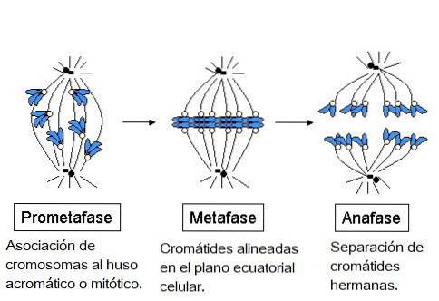
In the prometaphase, the formation of the mitotic machinery occurs, thanks to the assembly of the microtubules and their penetration into the interior of the nucleus. Sister chromatids linked by the centromeres are generated and these, in turn, bind to the microtubules.
During metaphase the chromosomes align in the equatorial plane of the cell. The spindle is organized into a central mitotic spindle and a pair of asters.
Each aster is made up of microtubules arranged in a star shape that extend from the centrosomes into the cell cortex. These astral microtubules do not interact with chromosomes.
It is then said that the aster radiates from the centrosome to the cell cortex and participates both in the location of the entire mitotic apparatus and in determining the plane of cell division during cytokinesis..
Later, during anaphase, the microtubules of the achromatic spindle are anchored by a positive end to the chromosomes through their kinetochores and by a negative end to a centrosome.
Separation of sister chromatids into independent chromosomes occurs. Each chromosome attached to a kinetochore microtubule moves toward a cell pole. Simultaneously, the separation of the cell poles occurs.
Finally, during telophase and cytokinesis, nuclear membranes are formed around daughter nuclei and chromosomes lose their condensed appearance..
The mitotic spindle disappears as the microtubules depolymerize and cell division occurs entering the interface.
The mechanism involved in the migration of the chromosomes towards the poles and the subsequent separation of the poles from each other is not exactly known, however; It is known that interactions between the kinetochore and the microtubule of the spindle attached to it are involved in this process..
As each chromosome migrates toward the corresponding pole, depolymerization of the attached microtubule, or kinetochoric microtubule, occurs. It is believed that this depolymerization can generate the passive movement of the chromosome attached to the microtubule of the spindle..
It is also believed that there may be other motor proteins associated with the kinetochore, in which the energy from the hydrolysis of ATP would be used..
This energy would serve to drive the migration of the chromosome along the microtubule to its end called "less" where the centrosome is located..
In unison, the depolymerization of the end of the microtubule that joins the kinetochore, or the “plus” end, could occur, which would also contribute to the movement of the chromosome..
The achromatic or mitotic spindle is a cellular structure that fulfills the function of anchoring the chromosomes through their kinetochores, aligning them with the cell equator and finally directing the migration of the chromatids towards the opposite poles of the cell before their division, allowing the distribution Equitable genetic material between the two resulting daughter cells.
If errors occur in this process, a lack or excess of chromosomes is generated, which translates into abnormal development patterns (to occur during embryogenesis), and various pathologies (to occur after the birth of the individual).
There is evidence that the microtubules of the achromatic spindle participate in determining the location of the structures responsible for cytoplasmic division..
The main evidence is that cell partition always occurs in the midline of the spindle, where the polar fibers overlap..
Evolutionarily it has been selected as a very redundant mechanism, in which each step is carried out by microtubule motor proteins.
It is believed that the evolutionary acquisition of microtubules was due to a process of endosymbiosis, in which a eukaryotic cell absorbed a prokaryotic cell that exhibited these achromatic spindle structures from the environment. All of this could have happened before the onset of mitosis.
This hypothesis suggests that the microtubule protein structures could have originally fulfilled a propulsion function. Then, when becoming part of a new organism, the microtubules would constitute the cytoskeleton and later, the mitotic machinery.
In evolutionary history there have been variations in the basic scheme of eukaryotic cell division. Cell division represented only some phases of the cell cycle, which is a major process.


Yet No Comments