
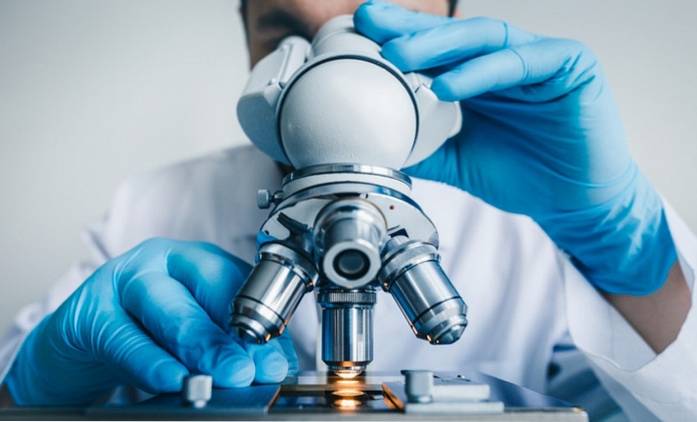
The importance of the microscope in medicine, health and science in general it is because it is a tool that allows observing cells, particles, bacteria and microbes, among other organisms and elements that would be invisible to the naked eye.
The microscope was created in the late 16th century by Zacharias Janssen. In its first design it had a pair of glass lenses, to generate increased vision. With the passage of time and the evolution of techniques, the electron microscope was reached, which allows us to see even the interior of a living cell.
The arrival of the microscope produced a revolution in the way of thinking of the human being, through which the body and its affections began to be studied in a scientific way, starting from the meticulous observation of the same.
Today, taking advantage of advances in technology, microscopes enable the detailed study of cells and molecules, among others, allowing specific research on drugs and diseases..

Since it was invented, the microscope has helped study organisms and particles, invisible to the naked eye, whose existence was not known. This has allowed the creation of new areas of study, both in biology, medicine and science..
In addition, it began a phase of experimentation and formulation of scientific theories, based on observations made with magnifying lenses. Making it possible to identify, for example, microorganisms that cause diseases, or even discover new, tiny living beings, of which there was no knowledge.
On the other hand, there are different types of microscopes, useful in various fields of study, such as medicine, health and natural sciences. Each of these fields has benefited from the use of the microscope, applied to its specific topics of interest..
Surgical microscopes are used to perform surgeries of different medical specialties, during which, due to the delicate nature of the tissues to be intervened, the surgeon needs to increase his vision..
In this way, the manipulation and repair of a large number of systems such as veins, blood vessels and nerves, is more precise and better results are obtained..
This type of microscope allows the surgeon to be in a comfortable position for handling the instruments, without worrying too much about handling the device, thanks to the fact that it can easily magnify the image of the desired sector..
Some of the medical fields where this type of microscope is used more frequently are ophthalmological, neurological and dental, among others..
The super-resolution microscope renewed optical microscopy, exceeding the resolution limit that was believed to be the maximum, bringing the limit of visibility to a nanometric scale, that is, one billionth of a meter.
It is for this reason that this microscope makes it possible to observe molecules found inside living cells..
The use of the super-resolution microscope is currently applied to the study of diseases such as Parkinson's and Alzheimer's.
Electron cryomicroscopy allows to obtain atomic precision when making observations of macromolecular structures and nanometric structures, without the need to use a large amount of sample volume.
In addition, thanks to advances in the area of image capture and data processing, three-dimensional models of the observed element can be obtained, which facilitate the interpretation of the images and help to better understand them..
Due to the fact that it does not need large quantities of samples, nor their crystallization, as was done previously, cryomicroscopy electron technology is widely used in the field of structural biology..
Another of the fields where it is used more frequently is that of medicine, allowing the three-dimensional construction of the parts that make up different types of cells. It is also a useful tool to study viruses such as HIV, facilitating the development of effective treatments for their eradication, based on its understanding and careful analysis..
This type of microscope is characterized by creating an electron beam, which is directed so that it impacts on a tissue sample to be observed, and when passing through it, generates a detailed image of it..
The image magnification scale is about one hundred thousand times that of the original sample size. Allowing, in this way, the visualization of the interior of cells and identifying DNA molecules, chromosomes and atoms.
It is for this reason that, through the use of this type of microscope, it is possible to investigate diseases, and develop drugs and treatments to combat them more effectively..
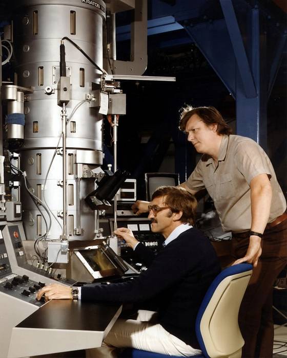
With an approximate height of 1.5 meters, and a weight of one thousand kilograms, this type of microscope is essential in the fields of medicine, the pharmaceutical industry, the materials industry, biology and nanoparticle analysis..
The tunnel effect microscope is commonly used in the field of nanotechnology, since it allows to visualize the atomic organization of the particles.
The operation of the microscope is based on the fundamentals of quantum mechanics, capturing electrons and giving way to the visualization of high quality images, where it is possible to see each atom separately. In addition, it has the possibility of obtaining images in three dimensions, and modifying the molecular composition of the substances observed..
Cleaning of surfaces, controlled vibrations and sophisticated electronics are necessary for its correct operation..
The fluorescence microscope is widely used in the field of biology, this is because this method is very specific and offers the possibility of observing a sample in detail.
Its operation consists of taking advantage of the fluorescent properties of the sample to be studied, to capture detailed images of it. For this, gas lamps are used, such as mercury vapor lamps, which emit a particular wavelength, which causes the sample to emit light under its influence..
With this type of microscope, the quantity, distribution and location of a molecule within a cell can be determined..


Yet No Comments