
The spinal cord is the main information pathway connecting the brain and the peripheral nervous system..
Contents
It is located in the vertebral foramen and is made up of 31 segments: 8 cervical, 12 thoracic, 5 lumbar, 5 sacral, and 1 coccygeal. One pair of spinal nerves exits from each segment of the spinal cord.
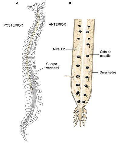
The length of the spinal cord is approximately 45 cm in men and 43 cm in women. The spinal cord is shorter than the length of the bony spinal column; the spinal cord extends downward only to the last thoracic vertebra. The nerves that extend from the spinal cord from the lumbar and sacral levels must run in the spinal canal for a distance before they exit the spine. This collection of nerves in the spinal canal is called the cauda equina (which means "horse's tail")..
It is very delicate and, therefore, has protection systems, among which the vertebral column stands out, which is made up of bones called vertebrae. Although the spine is somewhat flexible, some of the vertebrae in the lower parts of the spine are fused together.
It is also protected by the meninges and cerebrospinal fluid..
The spinal column is made up of twenty-four individual vertebrae that correspond to the cervical (neck), thoracic (chest), and lumbar (lower back) regions and the sacral and coccygeal vertebrae (in the pelvic area ).
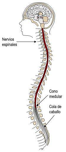
The spinal cord passes through the foramen of the vertebrae, from the first cervical vertebra (at the base of the skull) to the superior margin of the second lumbar vertebra and is therefore shorter than the vertebral column (it represents about 2/3 of the length of the column).
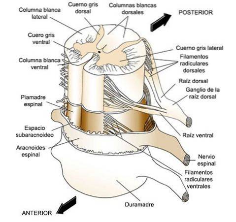
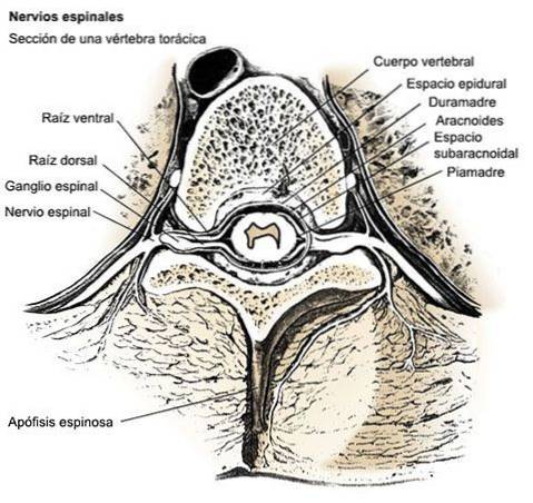
Like the brain, the spinal cord is covered by three layers of tissue (meninges). The spinal cord and meninges are contained in the spinal canal, which runs through the center of the spinal column..
Just as the skull protects the brain, the vertebrae protect the spinal cord. The vertebrae are separated by discs made of cartilage, which act as cushions, reducing the forces generated by movements such as walking and jumping..
The cord is surrounded laterally by the spinal nerves, axons of neurons that enter and leave the spinal cord and communicate with the rest of the body. Thirty-one pairs of nerves enter and leave the medulla, one for each side of the medulla:
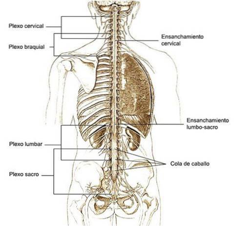
Each spinal nerve exits through the space between two vertebrae. In adults, the spinal cord is shorter than the vertebral column, therefore, in the lower segments of the cord the nerves must seek their exit at much lower levels, so that the lumbosacral roots form the cauda equina.
The cylinder formed by the medulla does not have the same diameter everywhere, but is thicker at the cervical level (cervical intumescence or dilation, innervation of the arms) and at the lumbosacral level (lumbar intumescence, leg innervation). These dilations correspond to regions of the medulla that innervate the extremities..
The spinal nerve roots are the bundles of fibers that come out of the spinal cord. For each spinal segment, (which is the area of the spinal cord that corresponds to the level of the vertebra through which the nerves exit to all parts of the body), there are four nerve roots: two in the front (ventral) and two behind (numbers).
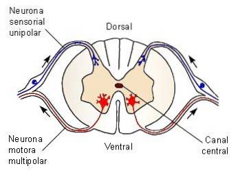
These roots, a right and a left, that is, one on each side of the spinal column, contain the nerves that control the movement of the body. The nerves and ventral nerve roots are called motor neurons.
They are efferent fibers that carry motor information from the spinal cord to the muscles.
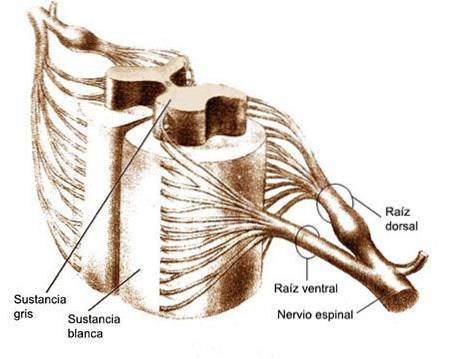 Dorsal roots
In the back, nerves (again one on the right and one on the left) carry sensory information from the body to the spinal cord or brain and are called sensory neurons. Once it reaches the spinal cord or brain, sensory information transmitted through sensory neurons is interpreted as sensation..
A dermatome is the area of the skin where the activity of a spinal or cranial nerve is received.
Dermatomes receive signals from sensory nerves in a spinal nerve root. A spinal nerve root is a mixture of several types of sensory, motor, and autonomic nerves that branch off from the spinal cord. The nerve root lies in an arch called the intervertebral foramen, which is a hole in the side of the spine made up of parts of individual vertebrae as they are stacked on top of each other..
Beyond the foramen, the nerve root begins to branch into individual nerves to reach and innervate all areas of the body.
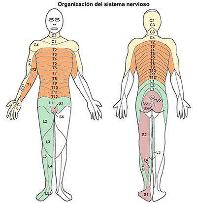
Therefore, in the medulla we can distinguish different spinal segments, each of which is linked to a spinal nerve. Actually, there is quite a lot of overlap in the innervation of adjacent areas.
The longitudinal grooves divide the spinal cord into the right and left halves. The ventral groove is known as the ventral medial fissure, and the dorsal groove is known as the dorsal medial groove..
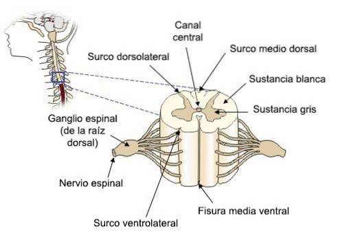
Cross sections of the spinal cord show us a clear division between white matter (exterior) and gray matter (interior).
The fundamental structure of the medulla is maintained throughout its entire length, although the proportions vary.
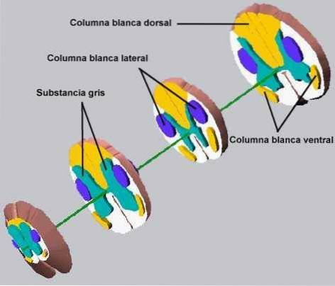
The gray matter is shaped like an H or a butterfly. In the middle there is a central channel, through which the cerebrospinal fluid circulates. It consists of ventral horns and dorsal horns, and an intermediate zone. At some levels (thoracic and upper lumbar), there is also a small lateral horn (efferent sympathetic neurons.)
The white matter is made up of nerve fibers, called axons, that run up and down the cord. Each group of axons carries a specific type of information that it needs to communicate. The ascending tracts of the axons communicate with the brain, while the descending tracts carry signals from the brain to various muscles and glands throughout the body..
Keeping in mind the gross anatomy and the internal organization of the cord, we can trace the path that sensory and motor information follow as they enter and leave the cord.
The main functions of the spinal cord include:
Electrical communication. Electrical currents travel up and down the spinal cord, sending signals that allow different segments of the body to communicate with the brain..
Control of the movement of the ambulation. During ambulation, the muscle groups in the legs are constantly contracting. The action of moving forward step by step may seem incredibly simple to us, as we have been doing it all our lives, but in reality there are many factors that must be properly coordinated to allow this movement to occur. These central pattern generators in the spinal cord are made up of neurons that send signals to the leg muscles, causing them to extend or contract, and produce the alternating movements that occur when a person walks..
Reflexes. These are predictable involuntary responses to stimuli involving the brain, spinal cord, and nerves of the peripheral nervous system (PNS)..

Bear, M.F .; Connors, B.W. i Paradiso, M.A. (1998). Neuroscience: exploring the brain. Barcelona: Masson-William & Wilkins Spain.
Bloom, F.E. i Lazerson, A. (1988). Brain, Mind, and Behavior. New York: Freeman and Company.
Carlson, N.R. (1999). Physiology of behavior. Barcelona: Ariel Psychology.
Carpenter, M.B. (1994). Neuroanatomy. Fundamentals. Buenos Aires: Editorial Panamericana.
Del Abril, A .; Ambrosio, E .; De Blas, M.R .; Caminero, A .; De Pablo, J.M. i Sandoval, E. (eds) (1999). Biological basis of behavior. Madrid: Sanz and Torres.
Delgado, J.M .; Ferrús, A .; Mora, F .; Rubia, F.J. (eds) (1998). Neuroscience Manual. Madrid: Synthesis.
Martin, J.H. (1998) Neuroanatomy. Madrid: Prentice Hall.
Nelson, R.J. (1996) Psychoendocrinology. The hormonal bases of behavior. Barcelona: Ariel.
Netter, F.M. (1987) Nervous System, Anatomy and Physiology. A Ciba Collection of Medical Illustrations (volum 1) Barcelona: Salvat.


Yet No Comments