Mexican Leishmania it is a Euglenozoa of the Kinetoplastea class. It is an obligate parasite that causes the disease known as leishmaniasis. In its life cycle it presents two completely different stages or body shapes, one of them elongated and flagellated and the other rounded or oval and lacking a flagellum..
In addition to the difference in shape, these two phases also differ in their habitat. The first one, known as promastigote, is extracellular and multiplies in the intestine of an insect vector; while the second, or amastigote, is intracellular and multiplies within human macrophages.

Leishmaniasis is a zoonotic disease that can have as reservoirs different species of mammals, generally dogs. It also uses blood-sucking mosquitoes, mainly of the genus Lutzomyia, as intermediate hosts and vectors. Apart from L. mexicana, there are other species of the same genus, all causing the disease.
Leishmaniasis can present in five clinical forms, localized cutaneous (LCL), recurrent (LR), diffuse cutaneous (LCD), mucocutaneous (CML) or spurious, and visceral (LV) or Kala-azar. Mexican Leishmania has been associated with localized and diffuse skin forms.
Article index
Mexican Leishmania it is a digestive parasite with an elongated and flagellate shape (promastigote) and a rounded and flagellate shape (amastigote). Additionally, there are several different forms of promastigotes that differ in their relative size and that of their flagellum..
The promastigote form is extracellular and reproduces only in the digestive tract of the intermediate host. While the amastigote form is intracellular and reproduces inside the macrophages of the definitive host.
The kinetoplast is made up of thousands of circular molecules and is located in front of the nucleus.
The genome of this species is made up of 34 chromosomes, with a fusion of chromosomes 8 and 29 and also chromosomes 20 and 36, thus presenting two fewer chromosomes than congeneric species distributed throughout Europe, Asia and Africa..
Mexican Leishmania has a complex life cycle, with a definitive host that is a mammal, including man, and an intermediate host represented by a blood-sucking insect.
Mexican Leishmania is taxonomically located in the phylum Euglenozoa, class Kinetoplastea, order Trypanosomatida and in the genus Leishmania. This genus was initially described by Borovsky in 1898, and the ordering of the species is not yet firmly established..
The criteria that prevailed for the original definition of the species of the genus were clinical, based on the type of leishmaniasis caused. The species were described Leishmania tropica, causing cutaneous leishmaniasis and Leishmania donovani, responsible for the visceral form of the disease.
Later geographic criteria prevailed. This allowed the description of new species up to a total of 39. In recent years, researchers have used molecular biology and phylogenetic tools to simplify the classification within the genus..
Mexican Leishmania is still considered a valid taxon, located within the subgenus Leishmania, along with the species L. donovai, L. major Y L. tropica. Seven species have been synonymised to L. mexicana, including L. amazonensis already L. venezuelensis.
The leishmaniasis parasite has two body forms: promastigote and amastigote:
It is considered the infective form. It is elongated and flagellated and has a size that will vary depending on the phase in which it is found:
The length of the body ranges from 6.5 to 11.5 µm. Another characteristic of this phase is that its flagellum is shorter than the body.
This phase is responsible for adhering to the microvilli of the epithelial cells. It has a length greater than 12 µm and in addition the flagellum is slightly shorter than the body.
The body length ranges between 6.5 and 11.5 µm, while the length of the flagellum is greater than that of the body.
It is the form that the insect transmits to the mammal when it bites it to feed. The size of the flagellum is still greater than that of the body, reaching less than 8 µm.
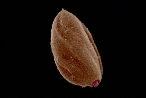
It constitutes the replicative form. It is round or oval with a diameter that ranges between 2 and 5 μm. Lacks scourge.
The life cycle, Mexican Leishmania It begins when an infected sandfly bites a mammal (including humans) for food. At that time, it injects metacyclic promastigotes into the skin of the mammalian host..
Promastigotes are phagocytosed by macrophages and dendritic cells. The parasites are not digested, but remain within a parasitophorous vacuole, where they transform into amastigotes and divide by fission..
The multiplication of the parasites causes the lysis of the infected cell, for which the amastigotes are released again to infect new cells and affect the skin tissues.
When an uninfected sandfly feeds on a diseased mammal, it ingests macrophages loaded with amastigotes and acquires the infection. The parasites reach the intestine in the form of amastigotes where they will transform into promastigotes.
Promastigotes go through each of the phases of this stage while dividing, until they transform into metacyclic promastigotes that migrate to the proboscis of the insect..
If in this phase the insect bites an uninfected mammal, it will inject the metacyclic promastigotes and a new cycle will begin..
Leishmaniasis is a disease caused by different species of Leishmania, and can affect the skin (localized, recurrent and diffuse cutaneous leishmaniasis), skin and mucous membranes (espundia) or internal tissues (visceral or Kala-azar).
Leishmaniasis, in any of its clinical forms, affects more than 12 million people worldwide. It is estimated that at least 2 million people are infected annually. Mexican Leishmania has been associated with only two of these clinical forms of the disease.
The main vectors of the disease are sandfly insects of the genus Lutzomia, which reach a maximum size of 4 mm.
This type of leishmaniasis occurs when amastigotes do not spread beyond the site of the bite, hence the name localized. Sandflies must feed in this area to acquire the parasite. It is the most common form of leishmaniasis. Can heal spontaneously.

It is a recurrent and disseminated infection that tends to reappear after treatment is finished. It does not heal spontaneously. The lesions that occur in this type of leishmaniasis are usually asymptomatic, with no tendency to ulcerate. It is a rare form of the disease.
Leishmaniasis can be asymptomatic or present different symptoms, depending on the clinical form, after an incubation period that can range from a week to several years, although the latter is rare..
The initial signs of the disease consist of the appearance of vascularized itchy papules in the area of the insect bite. Nodules or hyperkeratosis may also appear instead of papules.
Papules appear with raised edges, ulcerate, and may be dry or ooze after a few weeks, forming lesions that occur most frequently on the hands, feet, legs, and face. Injuries are not painful.
Lymph nodes may become swollen, although no elevation of body temperature occurs.
This type of disease occurs when amastigote spreads through the skin to other tissue and the lymphocytes are unable to react to antigens of Leishmania (anergy).
The main manifestations are thickening of the skin in the form of plaques, papules or nodules. There are no ulcers or additional symptoms.
For the diagnosis of the disease, the detection and identification of amastigote is necessary. This requires obtaining a skin sample by scraping or aspirating the lesion. The sample must then be stained with Giemsa stain to show and identify amastigote..
Cultures should be performed in NNN media for at least 4 weeks, as growth can be slow. The identification technique of the isolated species can be monoclonal antibodies, isoenzyme analysis, hybridization with DNA probes or polymerase chain reaction..
Serology is not recommended as it is not a very sensitive test in these cases..
There is no specific optimal treatment for the disease. Localized cutaneous leishmaniasis tends to heal spontaneously after several months and leaves scars. Treatment in this case helps to improve healing and prevent the spread of the parasite as well as relapses of the disease..
Traditional treatment consists of the use of antimonials such as sodium stibogluconate or meglumine antimoniate, administered intramuscularly or intralesionally. These drugs can have serious but reversible side effects, such as kidney failure, muscle pain, and liver or heart toxicity..
Recent treatment alternatives are amphotericin B, pentamidine, mitelophysin, paromomycin, thermotherapy and also chemotherapy.
Suggested preventive measures to avoid the disease include:
Attempting to reduce the size of vector populations by using insecticide spraying.
Treat insect screens, mosquito nets, clothing, and sheets with repellants with diethyltoluamide (DEET), permethrin, or pyrethrin.


Yet No Comments