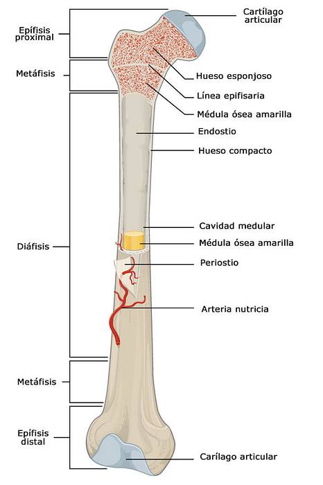
The metaphysis It is the area of the long bones that is located between the epiphysis (upper and lower part) and the diaphysis (central part). In the bones of children and adolescents who are in the growth period, the metaphysis is separated from the epiphysis by a transitional zone called growth cartilage, which allows bones to lengthen.
Once bone development is complete, around age 18 for women and age 21 for men, the growth plate calcifies and the metaphysis is permanently attached to the epiphysis. This union is known as epiphyseal line.

Histologically, this part of the bone is made up of trabecular or spongy bone tissue, that is, it contains bone marrow, which is responsible for the development of blood cells and their release into the stream..
The area of the metaphysis is richly vascularized and these blood vessels are responsible for supplying the growth cartilage that is close to it..
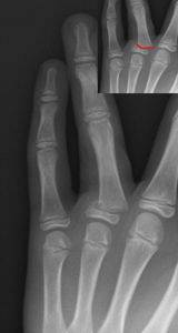
When metaphysis fracture occurs, the growth plate may be involved. This type of injury occurs in sports children or in the case of severe trauma.
Treatment is simple, but the diagnosis can go unnoticed, so the patient should be seen by a specialist if they have a long bone injury.
Article index
Long bones consist of three parts, the epiphyses that are located at the ends, the diaphysis, which forms the middle part of the bone, and the metaphysis that is located between these two portions..
The metaphysis is an area found in the long bones. During growth it is separated from the epiphysis by a specialized cellular cartilage, which is called growth cartilage..
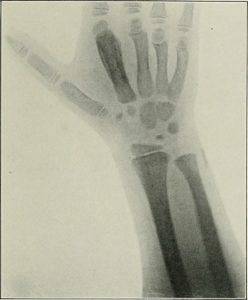
In large bones such as the femur, tibia or radius, there are two metaphyses. One at the top, or proximal, and one bottom or distal. Smaller long bones, such as interphalangeal or metacarpal bones, have a single metaphysis.
The bone tissue that forms the metaphysis is trabecular or spongy. This type of tissue withstands rebound impact well and transfers the vibrations from these impacts to hard or compact bone tissue. It also has an architecture that consists of small bony partitions within which is the bone marrow..
Blood cells are formed within the bone marrow that will be released into the circulation..
The metaphysis is a fundamental part of the bone that contains a complicated network of blood vessels that are responsible for nourishing the nearby cartilage.
The cells that will form the bones begin to differentiate from the 4thto gestation week, however, is not until 8to week that you can recognize an organized formation of what will be the skeleton.
Long bones originate between 9to and 10to week and its process begins with the formation of cartilaginous tissue around which cells that differentiate into osteocytes, or bone cells, are grouped.
The diaphyses are the first structures to calcify while the epiphyses and metaphyses have a more complex formation process.
The diaphysis is made up of bone tissue, but at its junction with the metaphysis, a cartilaginous tissue is formed that prevents calcification and adhesion between these parts..
The cartilage found in this area is a specialized tissue and has the property of lengthening with growth..
In long bones, the differentiation between the metaphysis and the diaphysis can be clearly observed through a conventional radiological study.
When the child is born, its skeleton is fully formed and the long bones have these sections of cartilage that will allow growth.
During the growth period, the bones are not completely calcified. This means that there are areas that are maintained with a tissue softer and more elastic than bone, which allows it to lengthen.
Between the epiphyses and metaphyses is this tissue called growth cartilage or growth plate.
The growth plate does not contain blood vessels. In young children it is nourished by the vasculature of the epiphysis, but in adolescents and adults the vascular network of the metaphyses is responsible for irrigating this area..
The irrigation is given in a 1) intraosseous way, through the channels that are formed inside the spongy tissue, 2) and extraosseous, by the blood vessels that are on the surface of the metaphysis.
This type of irrigation prevents the growth plate from losing its blood supply in the event of a trauma that injures the epiphysis..
Another important function of the metaphysis is to absorb the impacts of the joints and transfer them to the diaphysis, which is a stronger and more resistant bone tissue. In this way it prevents the joint complex from being overloaded.
Lesions of the metaphyses are especially important during the growth period. This is due to its relationship with the growth plate..
About 30% of fractures in growing individuals involve the growth plate and, from this percentage, it is extrapolated that 75% have a metaphysis injury.
Fractures of the metaphysis that involve damage to the growth plate are called Salter-Harris fractures. These are divided into five types, depending on the elements that are involved in the injury and its severity..
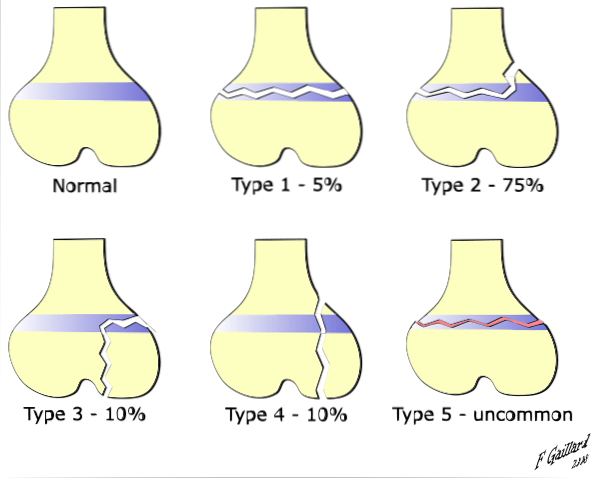
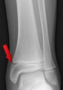
This type of fracture occurs in very active children, usually athletes. The most common are called Salter type II; are linear ruptures that partially separate the metaphysis and the growth plate of the epiphysis.
In some cases it is difficult to see them clearly on a conventional radiograph. The diagnosis is made by correlating the history with the physical examination and radiological findings..
Type II Salter fractures are easy to manage, with immobilization and rest, and do not interfere with the child's growth.
These lesions should be evaluated by a specialist, since when the timely diagnosis is not made, the patient can have repercussions on growth that are not reversible..


Yet No Comments