The microsporidia (Microsporidia) Is a phylum of fungi that groups more than 1400 species belonging to 200 genera. Its location in the Fungi Kingdom is controversial due to the absence of chitin in most stages of the life cycle, with the presence of chitin in cell walls being a widely used characteristic to define a fungus..
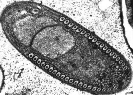
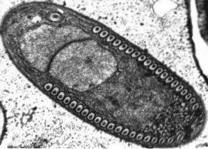
Microsporidia are eukaryotic cells. They have a well-defined posterior vacuole, nucleus, and plasma membrane. They are covered by a protective layer composed of proteins and chitin, which give it a high environmental resistance. They lack some typical eukaryotic organelles, such as mitochondria, the Golgi apparatus, and peroxisomes.
Microsporidia are obligate intracellular parasites of vertebrates and invertebrates. The most common species in the digestive system of humans are Enterocytozoon bieneusi Y Encephalitozoon intestinalis.
Human infection with microsporidia is called microsporidiosis. It occurs mainly in people who have undergone organ transplants or are immunosuppressed, such as those infected with the Human Immunodeficiency Virus. They also affect children, the elderly or people who wear contact lenses.
The genomes of the species of this phylum are used as models to study host-parasite interactions..
Article index
The fungi of the phylum Microsporidia form nonmotile spores that vary in size depending on the species. Spores measuring between 1 and 4 microns have been found in human infections.
The spores have several typical Microsporidia organelles:
The taxonomy and systematics of the phylum Microsporidia has changed over time and continues to be controversial. Initially it was classified in the Protista Kingdom, as a protozoan, due to the fact that they do not present chitin in the structures of most of the stages of the life cycle.
However, the results of studies using DNA techniques suggest that these organisms belong to the kingdom of fungi. Genomic data revealed that Microsporidia contain the genes necessary to produce chitin. In addition, chitin has been found in the resting spore structure..
There is also structural and metabolic evidence that allows Microsporidia to be recognized as true fungi. They apparently share a common ancestor with the phylum Zygomycetes and Mucorales..
The classification of this edge in terms of classes, orders and families is also controversial, so it continues to be reviewed and debated. Recent studies total about 150 genera and more than 1200 species.
14 species have been identified as disease producers in humans, distributed in the genera Anncaliia, Enterocytozoon, Encephalitozoon, Nosema, Pleistophora, Trachipleistophora and Vittaforma.
Microsporidia, in the form of spores, can survive in open environments for a long time and under adverse conditions. When spores enter the gastrointestinal tract of a host, they leave their active form. Mainly due to variations in the pH of the environment and due to variation in the cation / anion concentration ratio.
During the activation process, the cell expels the polar tube and penetrates the host cell membrane, injecting infectious sporoplasm into it. Once inside the cell, two key reproductive phases occur in the microsporidium.
On the one hand, reproduction occurs by binary fission (merogony) or multiple (schizogony). During this phase, the reproduction of cellular material occurs repeatedly before cell division occurs, producing rounded forms of multinucleated plasmodia (E. bieneusi) or multinucleated cells (E. intestinalis).
On the other hand, sporogony occurs, a process that gives rise to spores. Both phases can occur freely in the cytoplasm of cells or inside the gallbladder..
When the spores increase in number and fill the host cell's cytoplasm, the cell membrane ruptures and releases the spores to the surroundings. These mature spores, in their free state, can infect new cells, continuing the life cycle of microsporidia..
Microsporidiosis infections in humans are known as Microsporidiosis. Gastrointestinal tract infection is the most common form of microsporidiosis.
In the vast majority of cases, it occurs due to the ingestion of spores of Enterocytozoon bieneusi. Other times it can occur from infections of Intestinal Encephalitozoon.
Microsporidia spores are capable of infecting any animal cell, including insect, fish and mammalian cells. They can sometimes infect other parasites.
Some species have specific hosts. Encephalitozoon cuniculi harbors rodents, rabbits, carnivores, and primates. E. hellem in birds of the genus psittasis.
E. intestinalis in donkeys, dogs, pigs, cattle, goats and primates. Enterocytozoon bieneusi in pigs, primates, dogs, cats and birds. Annicaliia algerae stays in mosquitoes.
Infected animals and people release the spores into the environment with feces, urine and respiratory secretions. Thus, person-to-person infections may occur or contamination of water and food sources may occur, these being the most frequent sources of infection..
Infections by Enterocytozoon bieneusi Y Encephalitozoon intestinalis clinically manifest with watery diarrhea in immunocompetent adults and children, especially in people who reside or travel to tropical countries.
In immunocompromised patients, with HIV or another type of immune compromise, microsporidiosis presents as chronic diarrhea and wasting syndrome, cholangiopathy and acalculous cholecystitis.
Other species can cause urinary tract infection, hepatitis, peritonitis, encephalitis, urethritis, prostatitis, nephritis, sinusitis, keratoconjunctivitis, cystitis, cellulitis, disseminated infection, systemic infection, pneumonitis, myositis, and skin infection..
In patients with HIV infection, High Efficiency Antiretroviral Therapy (HAART) restores the immune response. It induces the elimination of the microorganism and the normalization of the intestinal architecture.
In most infections by microsporidia and especially by species of the genus Encephalitozoon Albendazole, a tubulin inhibitor, is used. The duration of treatment depends on the immune status of the patient and the type of infection, whether it is disseminated or localized..
Topical fumagillin is used in keratoconjunctivitis.
Immunocompetent patients can receive short treatments and sometimes the infection is overcome spontaneously, without the need for treatment.


Yet No Comments