
The erector spinae muscle It comprises a complex group of muscles, accompanied by tendons. All this is covered by a special connective tissue. This functional complex covers an important part of the back, mainly covering the lumbar, thoracic and cervical areas..
They are located in the middle area of the intrinsic muscles of the back. They have fascicles that run vertically along the spinal column. Each fascicle joins a structure, such as: skull, cervical, thoracic and lumbar vertebrae as well as at the level of the sacrum and ilium..
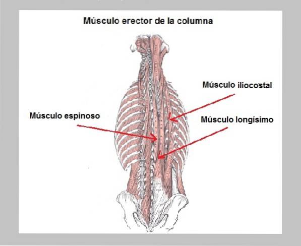
The complex group of erectors spinae is made up of three muscles, called iliocostal, longisimo, and spinous. These muscles are paired, that is, they are located on each side of the spinal column, specifically in the groove formed between the angles of the ribs and the spinous processes..
The erector muscle group is covered by a layer of connective tissue, called the thoracolumbar fascia, which encompasses the thoracic and lumbar region, while the cervical area is covered by the nuchal ligament..
Among the functions that this group of muscles and ligaments fulfill is to keep the spine in a straight or upright position, being called for this reason the extensor muscle of the spine. On the other hand, the spine is not a rigid structure, therefore, the set of muscles allows flexion movement.
Article index
The erector spinae muscle was known for a long time by the name of the sacrospinal muscle, a term currently in disuse. Today it is known as the erector spinae and is sometimes called the extensor spinae, due to the function it performs..
However, it is not a single muscle, therefore, it is considered a very important muscle complex. This is located in the middle part of the mass of muscles that are located on the back of the trunk..
Below the erector spinae muscle are the following muscles: intertransverse, multifid, rotator, and interspinous. While, above this are: the trapezius, the rhomboids, the latissimus dorsi, the serratus posterior, the quadratus lumbar and the angular shoulder blade..
The erector spinae group is made up of three muscles that are paired. These are located symmetrically on each side of the spine vertically. From the bottom up it can be said that the muscle complex extends from the pelvis to the skull. The muscle looks like a thick, broad band.
There are three muscles and they are called: spinous, long and iliocostal.
It is located right next to the spine (medial line of the body).
It is located in the middle, between the spinous muscle and the iliocostalis (intermediate line).
It is the outermost of the three and the furthest from the spine (lateral line of the body). It is divided into three regions according to the site where its fibers are inserted: lumbar, thoracic and cervical iliocostal.
It has already been mentioned that the large muscle complex comprises 3 muscles, but there are also ligaments and the thoracolumbar fascia. Therefore, the whole of this structure is divided by zones.
The spinous muscle and the longis muscle participate in this region. These cover the base of the skull, which in turn is covered by the nuchal ligament. Some authors call this part the erector cervical spine muscle..
All three muscles participate in this area: spinous, longus and iliocostal. They are seen as 3 pillars (from T12 to L1). The fibers of these muscles are thickest towards the base and finest towards the top. This zoneit is also known as the erector spinae muscle.
In this part, the separation of the three muscles is not distinguished, therefore, it appears as a single thick muscle mass. This area is also calledlumbar erector spinae muscle.
This region is basically covered by much finer tendons or ligaments, culminating in the shape of a point. This part the structure is finer or narrower. Corresponds to the common site of origin of the erector spinae muscle complex.
Its origin occurs at the level of the aponeurosis of the muscle under study. It corresponds to the common origin of the erector spinae muscle. This area has several insertion sites that are: iliac crest (upper third), sacrum (posterior part), spinous processes of the lumbar region and the sacroiliac ligaments.
In this area the 3 muscles are inserted, serving the spinous processes as the attachment site for the spinous muscle fibers. Whereas, the transverse processes serve as an attachment site for the muscle fibers of the longis. While, in the ribs the thorny is inserted.
In this area, the spinous processes also serve as an attachment site for the muscle fibers of the spinous bone, but it also inserts at the base of the occiput..
Likewise, the transverse processes continue to serve as a site of attachment for the muscle fibers of the longis muscle and it also inserts into the mastoid process of the temporal bone..
The complex group of muscles is innervated by the spinal nerves, specifically receiving lateral branches that come from the posterior branch.
Because it is a large muscle and has a long history, it is irrigated by many blood vessels depending on the area..
In the cervical area, it is supplied by the superficial and deep descending branches of the occipital artery, the transverse cervical artery, the deep cervical artery and the vertebral artery..
The dorsal or thoracic area is supplied by the dorsal branches of the superior, posterior and subcostal intercostal arteries..
And the lower or lumbosacral part is nourished by the dorsal branches of the lateral and middle sacral arteries.
Venous return is carried out under the same pattern. Changing the word artery for vein.
Its bilateral action is to extend both the neck and the spine, being essential to maintain a totally straight or upright position. In this sense, its action is concentric.
In its unilateral action, it fulfills a flexor function, allowing the movement of the spine and neck to one side or the other, depending on the muscle that is acting (right and left). The movement is executed to the same side of the muscle that is in action.
It also participates in the forward movement of the spine, where the erector spinae muscle plays a fundamental role in controlling descent, acting eccentrically..
Back pain is a very common ailment and most cases are muscular in origin. Pain may occur from muscle stretching or spasms and trigger points may appear.
The muscle fibers of the erector spinae can tear or stretch due to an excessively poorly balanced load on the back. This causes the muscles to become overloaded..
When the body is not warmed up before exercising certain exercises, cramps can occur, which is characterized by muscle contractions that are usually painful, affecting the deterioration of its function..
The muscle can suffer contractures at any given time, due to poor posture, muscle weakness, weight overload, among others. Trigger points cause pain, generally occurring at the lumbar level (unilateral), but can radiate to the gluteal area.
To relieve this muscle it is recommended to rest the first days of pain, it is also favorable to place heat in the affected area to increase blood flow.
Likewise, it is advisable to perform exercises, especially stretching, as well as to avoid being constantly in the same position, either sitting or standing. Finally, it is useful to perform physiotherapy (massage, exercises, electrotherapy, etc.)
In case of acute pain that does not stop with the aforementioned, there is a surgical alternative that eliminates the pain at its roots. This is achieved by blocking the erector lumbar spine plane..
To palpate the muscle, you must first locate it. A quick and easy way to do this is to tell the patient to lie on their stomach and then try to move their head, pelvis, and arms back. There you can see how the muscles are tense on either side of the spine. Once located, they can be palpated and massaged.
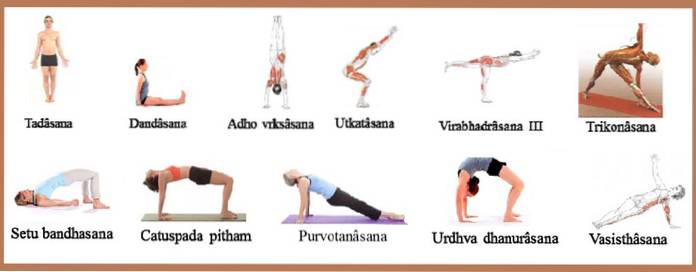
There are a variety of positions used in yoga that help strengthen this muscle. See the following figure.
Patients with this disorder have a deviated spine, which can be painless or painful. Pain is associated with chronic myofascial trigger points. One of the muscles most affected in this disorder is the erector spine muscle..


Yet No Comments