
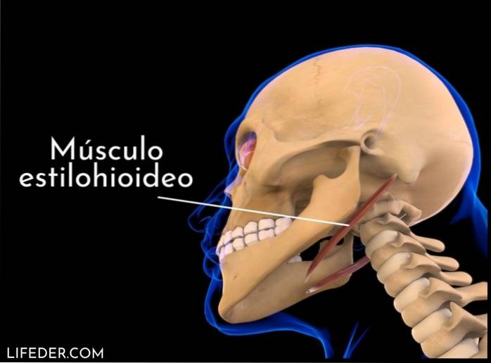
The stylohyoid muscle It is a small, thin, bilateral muscle that is located in the neck and extends in front of and above the digastric muscle. Due to its location, it belongs to the anterior group of neck muscles, topographically divided into muscles of the deep plane and the superficial plane..
The muscles of the superficial plane are separated by the hyoid bone into a suprahyoid group (those located above the hyoid bone), and an infrahyoid group (located below the hyoid bone). There are four muscles in the suprahyoid region: digastric, stylohyoid, mylohyoid and genihoid..
The function of this group of muscles is to lower the jaw by contraction, supported by the counterpart of the infrahyoid muscle group, thus allowing the hyoid bone to balance. Its name reveals its origin and insertion, since it originates in the styloid process and inserts in the lateral area of the body of the hyoid..
It is part of the muscular and ligamentous group called the Riolano branch, which is made up of the stylopharygeal, styloglossal, stylohyoid muscles and the stylomaxillary and stylohyoid ligaments, all of which are inserted into the styloid process of the temporal bone in the form of a bouquet..
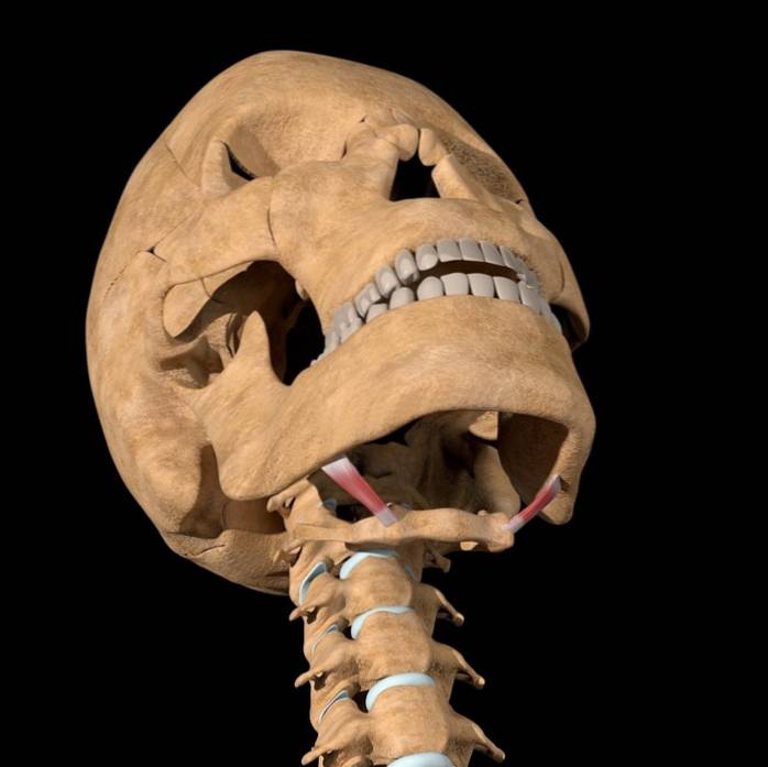
Its lower end divides to form an eyelet that allows the common tendon of the digastric muscle to pass through, giving it a unique characteristic among the muscles of the neck. The stylohyoid muscle draws the hyoid bone back during swallowing and lengthens the floor of the mouth.
It is located or has its origin in the posterior and lateral surface of the styloid process, near the base; passing downward and forward, it inserts into the body of the hyoid bone, at the junction between the body and the greater horn.
The styloid process is a pointed part of the temporal bone of the skull, which is located just below the ear and functions as an anchor point for a number of muscles. The stylohyoid muscle is usually divided near its attachment by the digastric tendon.
It accompanies the posterior belly of the digastric muscle throughout its entire trajectory, standing laterally and then behind it.
Medially it is related to the styloglossus muscle, from which it is separated by a space occupied by the external carotid artery from the retroestylar region to the parotid region..
The facial artery passes below the posterior belly of the digastric muscle and the stylohyoid muscle, and penetrates above these to the submaxillary cell.
The stylohyoid muscle initiates the swallowing action by pulling the hyoid bone in a posterior and superior direction; that is, it retracts and elevates the hyoid bone.
Elevate the tongue and lengthen the floor of the mouth; therefore it is considered that it helps swallowing and elevates the larynx.
The vascularization of the stylohyoid muscle is given by various arteries and arterioles of the external carotid artery according to its surface. For this, the stylohyoid muscle is usually divided into three thirds.
The first and second third receive blood supply from a pair of arterioles of the posterior auricular artery, which is a branch of the external carotid artery.
The third third is supplied by the hyoid branch of the lingual artery, which in turn is a collateral branch of the external carotid artery.
The stylohyoid muscle grows from the second pharyngeal arch and, therefore, its innervation is supplied by the facial nerve (VII cranial nerve), which is a mixed cranial nerve..
The facial nerve exits the skull through the stylomastoid foramen, supplying nerve branches to the digastric and stylohyoid muscles. They can be independent branches or through a common trunk.
The stylohyoid muscle branch can be inconsistent, not because of its presence or absence, but because of the difficulty in dissecting it, since it is very small and its location is complex and never exact..
Few conditions can affect this muscle. However, if the nerves surrounding the stylohyoid become irritated or damaged, the result can be chronic pain in the face, neck, and head..
Although there is no known cure for it, the prescription of non-steroidal anti-inflammatory drugs (aspirin and ibuprofen) can reduce pain and inflammation. In very severe cases, injections are used to numb the affected area..
Although both muscles have the similarity of suspending the larynx, they have some differences between them:


Yet No Comments