The neck muscles They are the muscles that help us move our head in different directions with respect to the rest of our body. They also assist us when we breathe and when we swallow, and allow us to keep our heads up, while promoting the continuous flow of blood to the brain..
The neck is the part of the human body that connects the skull with the torso. In many textbooks it is also defined as the beginning of the spinal cord and spinal column..
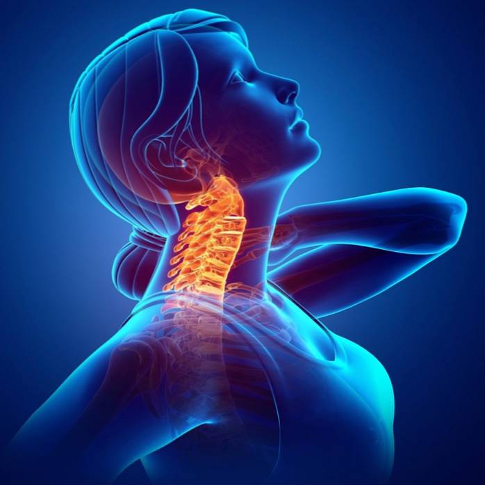
The vertebral column is the part of our axial skeleton that supports the weight of the head, protects the spinal cord (made up of the nerves that communicate the brain with the other organs) and is the site of attachment for the ribs and muscles of the back and neck.
This structure, made up of 24 bones called vertebrae, It is more or less 70 cm long in an adult human being and, in the neck, are the first 7 bones, which are the smallest and are also known as cervical vertebrae.
The neck is the part of our body that connects the head with the torso. It is formed, from the bone point of view, by the first 7 vertebrae of our spine, also known as cervical vertebrae
The neck performs important functions for our body, not only because it supports our head, but also because it allows us to speak, sing, chew and swallow, breathe, look to the sides, among others. Functions that, in part, are possible both due to the bone structure of the neck and its muscular components.
We can say that the neck is anatomically divided into three segments or regions:
An anterior or frontal.
Two sides (on each side).
A posterior.
Let's see which are the muscles that we find in each one:
Muscles of the anterior region
They are the muscles that are in the frontal region of the neck, that is, the region that is in the same plane as our mouth, our chest and our eyes.
These muscles, then, cover the anterior part of the neck and are divided into three groups, two of which owe their classification to their location with respect to the hyoid bone, which is a bone located more or less at the level of cervical vertebra number 3. , just below the jaw.
Here are the muscles of the anterior neck region:
Superficial muscles of the anterior region
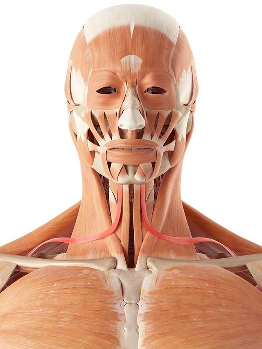
The muscles of the anterior region of the neck are those that are in the same plane as our face and our chest. They contribute to different processes of our daily life such as speech, breathing, chewing and swallowing, etc..
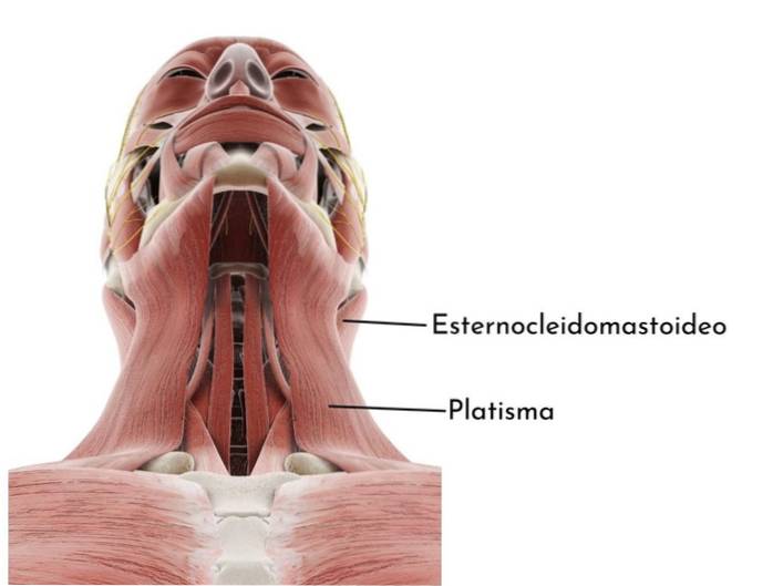
They are the most superficial muscles in this region of the neck, we can feel them with our hands if we stretch the neck a little, raising the chin. Two muscles belong to this group: the platysma and the sternocleidomastoid..
Platysma: it is a more or less flat muscle, shaped like a leaf or blade; it is located between the skin of the anterior neck region and the covering of the cervical vertebrae. It is born beyond the clavicle and covers the entire length of the neck, inserting into the jaw, the skin of the lower part of the face, the lower lip and the ends of the mouth.
Sternocleidomastoid: it is a long bone with two heads, one that connects with the clavicle and the other with the sternum; this muscle runs diagonally towards the mastoid bone, of the temporal bone of the skull.
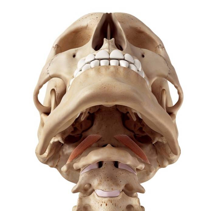
Suprahyoid muscles of the anterior region
There are four muscles found in the region superior to the hyoid bone. They are responsible for connecting this bone with the jaw and the base of the skull.
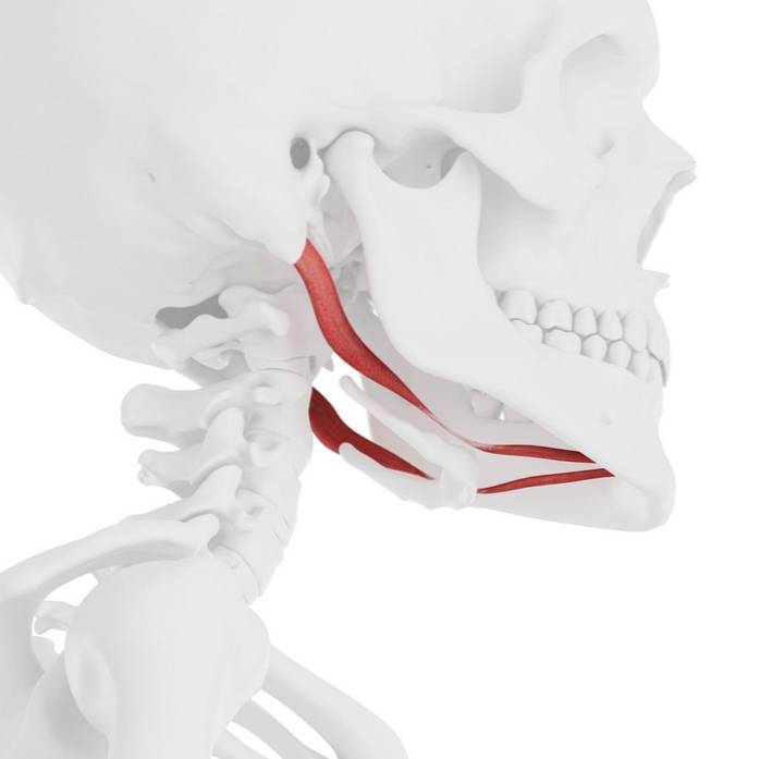
Digastric muscle: it is a small muscle that is under the jaw and goes from the mastoid bone in the temporal bone of the skull to the chin.
Digastric muscle
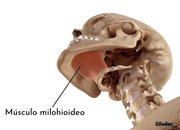
Mylohyoid muscle: is a flat muscle that forms the flat from the mouth; It arises on the inner surface of the mandible and attaches to the top of the hyoid bone. Helps us open our mouths and swallow.
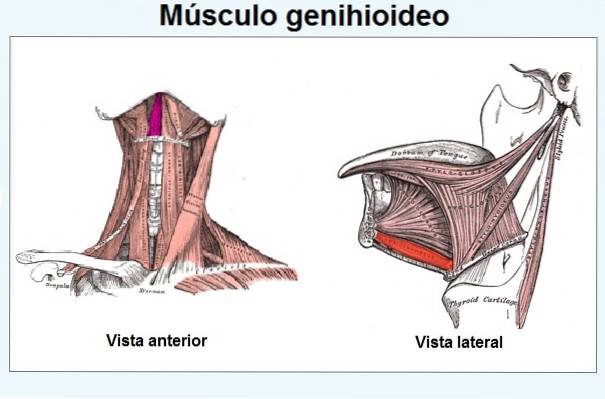
Muscle geniohyoid or geniohyoid: this muscle arises in the lower part of the jaw (in the chin area) and inserts longitudinally at the upper edge of the hyoid bone.
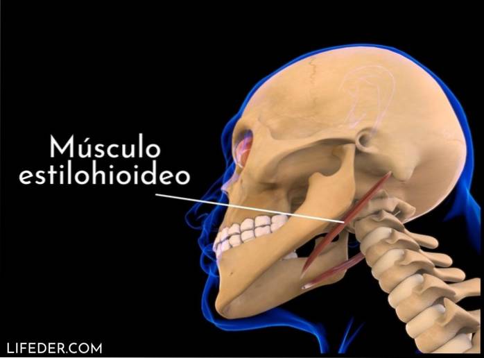
Muscle estilohyoid: extends between the temporal bone and the hyoid; facilitates elevation of the hyoid bone and retraction of the tongue.
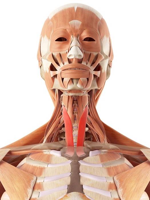
Infrahyoid muscles of the anterior region
They are the muscles found in the region below the hyoid bone. There are also four and they connect the hyoid with the larynx, sternum and scapulae, favoring chewing, swallowing (when we swallow) and vocalization (when we speak).
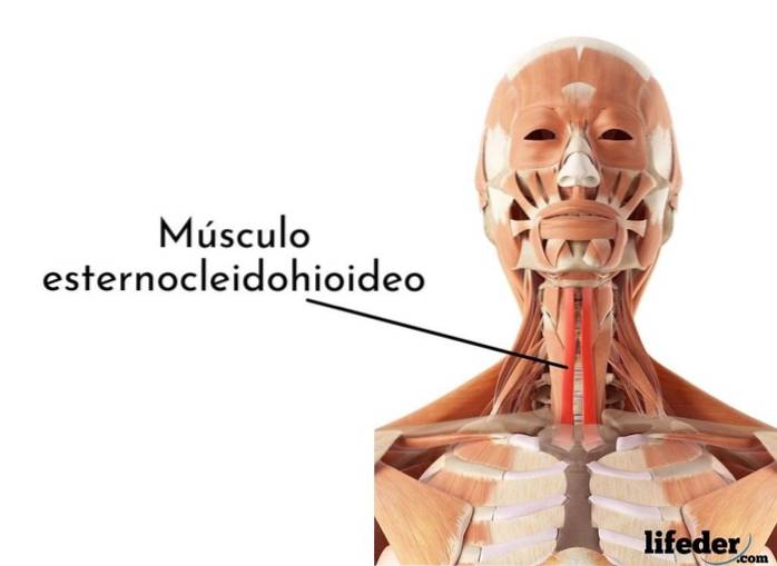
Sternocleidohyoid or sternohyoid muscle: is a ribbon-like muscle found in the region triangular By the neck. It arises in the upper part of the manubrium, in the sternum, and in the final middle part of the clavicle, inserting itself at the lower edge of the hyoid bone. Facilitates breathing and swallowing.
Location of the sternocleidohyoid muscle
Omohyoid muscle: it is an elongated, thin muscle, responsible for connecting the hyoid bone with the scapulae.
Omohyoid muscle
Muscle esternothyroid: is another ribbon-like muscle that is also found in the muscle triangle By the neck; arises from the cartilage of the first rib and inserts into the thyroid cartilage line.
Sternothyroid muscle
Muscle tiroids: it is an elongated muscle, also located in the muscular triangle of the neck and that arises in the lamina of the thyroid cartilage, inserting in the lower border of the greater horn of the hyoid bone.
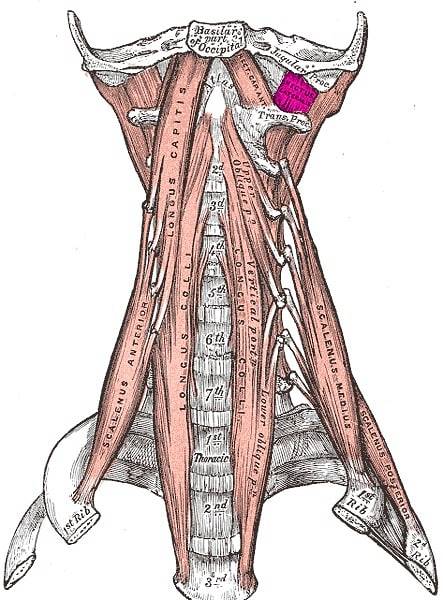
Muscles of the anterior vertebral region of the neck
They are the deep muscles of the anterior region of the neck. Their name indicates that they are located just in front of the cervical vertebrae of the spine that make up the neck. Its main function is to achieve the bending movement of the head.
Rectus anterior muscle of the head: it is a flattened muscle that arises on the lateral surface of the first vertebra and attaches to the occipital bone.
Rectus anterior muscle of the head
Lateral rectus muscle of the head: it is a small muscle that arises on the surface of the first vertebra and ascends to insert itself on the lower surface of the occipital bone.
Lateral rectus muscle of the head
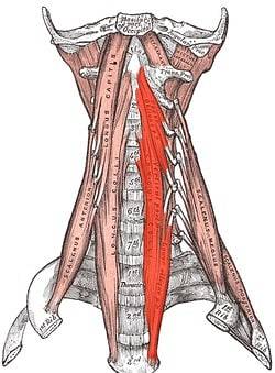
Long head muscle: it is an elongated flat muscle that arises between vertebrae 3 and 6, later inserting itself on the lower surface of the occipital bone; It is made up of four thin muscle "strips".
Long head muscle
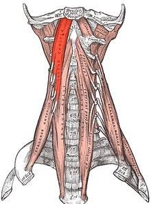
Long neck muscle: it is a very long muscle, as it covers the entire length of the cervical vertebrae that make up the neck. It is divided into an upper, an intermediate and a lower part, which are born and are inserted in different regions of the cervical region.
Long neck muscle
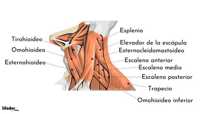
Muscles of the lateral region
They are the muscles found on the two left and right sides of the neck. These muscles allow lateral flexion of the neck and pass obliquely from the cervical vertebrae to the first two ribs.
Anterior scalene muscle: arises between vertebrae 3 and 6 and inserts on the upper border of the first rib.
Middle scalene muscle: is the longest of the three; arises between cervical vertebrae 1 and 2, and also between 3 and 7, inserting at the upper edge of the first rib.
Posterior scalene muscle: it is the shortest, as it arises in vertebrae 4 and 6 and inserts on the external surface of the second rib.
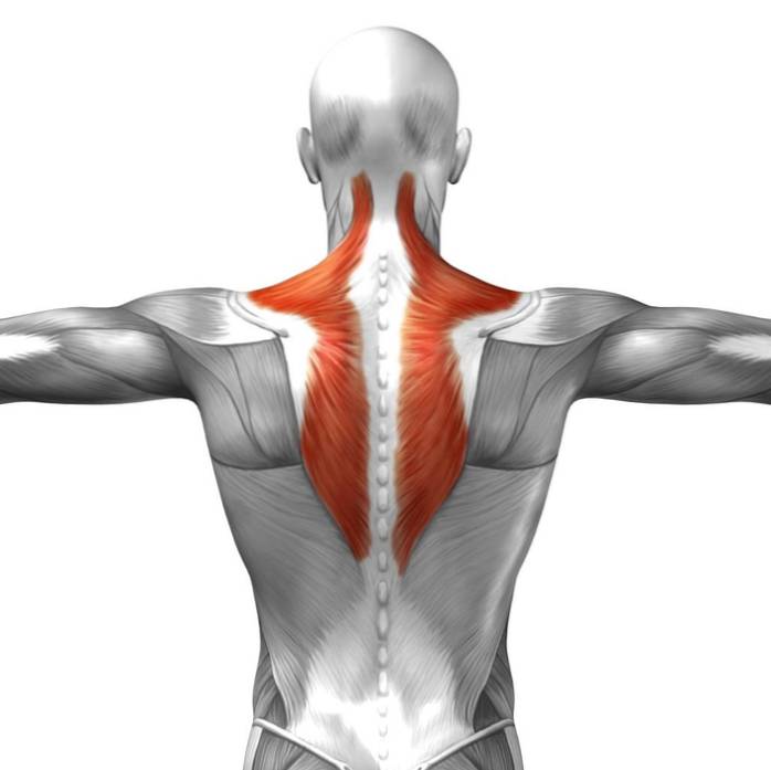
Muscles of the posterior region
Muscles in the posterior region stabilize the head and help perform various bending movements
They are the muscles that connect the skull to the spinal column and pectoral girdle. They are divided into three layers: a superficial one, a deep one and a very deep one. These muscles facilitate the lateral and contralateral rotation of the head and, in addition, participate in the movement of the scapulae.
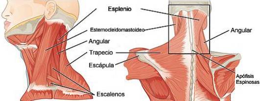
Surface layer: formed by the trapezius, splenius muscle and splenius muscle of the neck.
Deep layer: formed by the transversuspinous muscles which are the semispinatus muscle, the cervical semispinatus muscle and the cervical multifidus muscle.
Very deep layer: formed by the suboccipital muscles, the interspinous muscles of the neck and the intertransverse muscles of the neck.
References
Hamilton, W. J. (1982). Textbook of human anatomy. Springer.
Netter, F. H. (2014). Atlas of human anatomy, Professional Edition EBook: including NetterReference. com Access with full downloadable image Bank. Elsevier health sciences.
Putz, R., & Pabst, R. (2006). Sobotta-Atlas of Human Anatomy: Head, Neck, Upper Limb, Thorax, Abdomen, Pelvis, Lower Limb; Two-volume set.
Sendic, G. 2021. Muscles of the neck: An overview | Ken Hub. [online] Ken Hub. Taken from kenhub.com

Yet No Comments