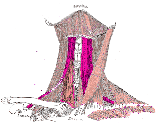
The infrahyoid muscles They are a muscle group made up of four thin, flattened muscles that lie below the hyoid bone. These muscles are located away from the spine, in front of the thyroid gland and the laryngotracheal canal..
To be able to locate the infrahyoid muscles through the palpation method, one must begin by placing the fingers on the lateral edge of the trachea, from there sliding slightly towards the sternocleidomastoid and following the different fibers of the muscles between the hyoid bone, the sternum , clavicle and scapula.
The patient will need to swallow to make sure that the location we locate is correct.
Article index
These muscles contribute to the mandibular descent when the oral cavity is opened. They are located in two planes: the superficial plane, made up of the sternohyoid and omohyoid muscles; and the deep plane, made up of the sternothyroid and thyrohyoid muscles.
Regarding innervation, the infrahyoid muscles present a common innervation as a fundamental characteristic; that is, they share the same origin, the upper root of the cervical loop.
The upper root of the cervical loop meets the lower root of the cervical loop and forms the hypoglossal loop. The nerves of the sternohyoid, omohyoid and sternothyroid muscles originate from the hypoglossal loop, while the nerve that goes to the thyrohyoid muscle is a direct branch of the hypoglossal nerve.
This muscle, also known as the sternocleidohyoid muscle, is the most superficial muscle of the infrahyoid muscles. It has a ribbon shape between 15 and 25 millimeters wide, its length goes from the upper extremity of the thorax to the hyoid bone.
It originates with a thick tendon that inserts into 3 different structures. Covers the posterior border of the clavicle, the posterior aspect of the sternoclavicular ligament, the lateral half of the manubrium of the sternum, and the first costal cartilage.
From there it travels upward to attach to the lower border of the hyoid body. This is covered below by the sternocleidomastoid and above by the omohyoid. It is superficial and medial.
The omohyoid muscle, also known as the omoplatohyoid or scapulohoid, is long and thin.
It is a digastric muscle; that is, it consists of two bellies: an upper one and a lower one. It also has an intermediate tendon that runs obliquely across the lateral cervical region, connected to the clavicle and the first rib..
The lower belly originates from the upper edge of the scapula. Medial to the scapular notch, ascends cranio-medially and merges into an intermediate tendon at the level of the lateral cervical region.
The intermediate tendon is connected to the carotid sheath, which surrounds the neurovascular bundle (including the common carotid artery, internal jugular vein, and vagus nerve)..
The upper belly of the muscle is detached from the medial tendon and is directed almost completely vertically, to attach to the lower and lateral border of the hyoid.
An important characteristic of this muscle is the relationships it has with various regions. Among these, the relationship with the posterior region of the neck stands out, where it is related to the scapular region; the lateral region, where it relates to the brachial plexus; and the carotid region and the anterior region of the neck, where it relates to the thyroid gland and larynx.
Its function is to depress the hyoid bone and the middle cervical fascia. It is a superficial and lateral muscle.
This muscle runs from the breastbone to the edge of the thyroid cartilage. It has its origin in the manubrium of the sternum, the most superior part of the sternum on its back side. From there it begins a short and vertical journey up.
It inserts in the oblique line of the anterolateral aspect of the thyroid cartilage and in the tubercles that limit the external aspect of the thyroid cartilage.
The sternothyroid muscle is shorter and wider than the sternohyoid muscle and lies below the latter.
The main function of this muscle is to depress the larynx for chewing and swallowing. This rise and fall of the larynx can also affect vocal range due to the ability to control pitch and volume..
The thyrohyoid is a short, flat muscle that looks like a continuation of the sternothyroid muscle. It arises from the thyroid cartilage of the larynx and ascends to join the hyoid bone. Its location in relation to the neck muscles is deep and lateral.
It originates from the anterolateral aspect of the thyroid cartilage and the tubercles that limit it; from there it goes upwards in a vertical direction, to insert on the edge and superficial face of the body of the hyoid.
Some of its fibers are also inserted at the base of the greater horn of the hyoid bone, in this way its contraction depresses the hyoid.
If the hyoid bone is fixed by the suprahyoid muscles, it can elevate the larynx. It has its innervation in the anterior branch of C1, transported within the hypoglossal nerve. It is innervated by the first cervical nerve, which joins the hypoglossal nerve for a short distance.
The infrahyoid muscles are responsible for fixing and lowering the hyoid bone and larynx when swallowing occurs (swallowing food, liquid or saliva) and contribute to phonation.
They also participate in the bending of the head. The infrahyoid muscles favor the lower jaw when the mouth is opened; fix the hyoid bone for the action of the suprahyoid muscles.
One thing to consider is that the sternohyoid, the sternothyroid and the thyrohyoid contribute to the structuring of the tracheostomy rhombus, the site of choice for access to the trachea.


Yet No Comments