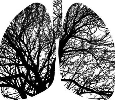
The pneumatocele to the pathological formation of a cavity within the lung parenchyma that fills with air. This cavity or cyst has very thin walls and sometimes, in addition to air, it can have fluid inside it. It is often confused with bulls, but these are not transitory as pneumatocele can be.
The etymology of the word, as in most medical words, has Greek roots. The first half, pneumon which means "lung" or "air", and the second part of the word comes from Kele, which has different meanings, including "tumor" or "herniation." The definitive term would be "air tumor" or "lung tumor".
Some classic medical texts describe cases of pneumatocele outside the lung. How can this be explained? The strict meaning of the word, according to certain authors, is "air cyst", so any tumor filled with air, wherever it may be, can be named that way. Hence, we speak of cerebral, intestinal or even cutaneous pneumatocele.
At present, the term pneumatocele has been dedicated almost exclusively to pulmonary pathologies. Respecting these scientific criteria, the development of this article is carried out explaining only the pulmonary pneumatocele. Some of the symptoms, causes and treatments associated with this pathology are mentioned below..
Article index
It is not surprising that the main symptoms of pneumatocele are related to the respiratory sphere. However, they are not limited to this device, since there are systemic or specific clinical manifestations in other organs..
Pneumatoceles are often asymptomatic. This will obviously depend on the size of it and its cause. When due to its characteristics it is capable of generating clinical manifestations, these occur due to the displacement of the structures around it or due to the compromise in gas exchange or in the ventilatory pattern.
Typical pneumatocele symptoms that involve respiratory anatomy and physiology include:
Although very nonspecific, respiratory distress is one of the typical signs of pneumatocele. It can be evidenced as an increase in respiratory rate, greater effort during inspiration, use of accessory respiratory muscles (intercostals), greater opening of the nostrils and panting.
When pneumatocele affects the junction between the alveoli (functional portion of the airway) and the pulmonary blood vessels, the exchange of gases between the body and the outside is disturbed. This is reflected in a decrease in the amount of oxygen in the blood, accompanied by an increase in carbon dioxide..
Clinically, distal and perioral cyanosis is evidenced. The fingertips and around the mouth turn purplish or bluish in color, and the blood becomes very dark. This phenomenon often goes hand in hand with dyspnea. Both signs are generated by the greater need for oxygenation that the body has.
If the pneumatocele is located on the periphery of the lung, close to the pleura, there may be pain. This is because one of the layers of the pleura is richly innervated and when pressed or pushed it hurts.
The intercostal nerves may also be affected, which in addition to causing pain can modify the respiratory pattern.
Due to the location of the pneumatocele, there may be involvement of the mediastinum, which would generate cardiovascular alterations. It is important to remember that the heart has a close anatomical relationship with the lungs, especially the left, and any injury that takes up space near the lungs can also affect it..
Mediastinal displacement caused by pneumatocele pressure is of more radiological than clinical significance. This means that despite the displacement being very evident on radiological studies, the symptoms are not as significant. However, there may be arrhythmias, dyspnea due to displacement of the trachea, or cyanosis..
Pneumatocele may also be associated with pericardial disorders. Depending on the cause, especially infectious or oncological, pericardial effusion and heart failure can occur. The patient will manifest chest pain, dyspnea, and weakness. The physical examination will show hypotension, paleness, and profuse sweating..
The causes of pneumatocele can vary a little between the different age groups, but in percentage terms they are almost always the same, among which the following are known:
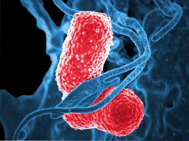
Infections appear to be the leading cause of pneumatoceles in both adults and children. The difference lies in the germ involved. In children, pneumatocele is more common as a complication of staphylococcal pneumonia, while in adults and immunocompromised patients, it is tuberculosis.
Chest trauma is another common cause of pneumatocele. For this to happen it is necessary that there is a laceration in the lung. The air will escape through this lesion but will be retained in the surroundings thanks to the rest of the structures of the thorax, thus favoring the appearance of the gas cyst.
Another risk group for the development of pneumatoceles is that of patients subjected to assisted ventilation for different reasons. This is due to barotraumas, or airway injuries caused by the pressure generated by the ventilator within the airways..
If, in addition, there is permanent communication between the airways and the lung parenchyma, or fistula, the pneumatocele can be perpetuated.
Aspiration of hydrocarbons or caustics, common in children, can cause injury to the trachea or bronchi and lead to a pneumatocele. Pulmonary infarcts have also been associated with the appearance of these lung cavities, as well as some oncological diseases such as lung cancer, breast and thoracic lymphomas..
The management of pneumatocele will depend on its origin. When associated with infections, the administration of antibiotics is necessary. Antimicrobials that attack staphylococci, such as oxacillin or vancomycin, are frequently indicated. Antituberculous chemotherapy is also essential when this is the cause.
Many pneumatoceles, especially those associated with pulmonary infections or those of idiopathic cause, can regress spontaneously. Conservative treatment is indicated when symptoms are mild or absent and pneumatocele was an occasional finding..
Surgery is the treatment of choice when there is a fistula that does not allow the pneumatocele to heal or when the respiratory symptoms are very severe. In these cases, the cyst must be removed in its entirety and repaired nearby damage that may cause its reproduction or reappearance.


Yet No Comments