
The neurohypophysis, Also called the posterior lobe of the pituitary or posterior pituitary, it is a structure that is responsible for storing and releasing two hormones: vasopressin and oxytocin. These hormones regulate the secretion of water, and the mammary glands and uterine contractions, respectively..
This structure is part of the pituitary gland or pituitary, belonging to the endocrine system. It is mainly composed of axons without myelin from the hypothalamus and blood capillaries.
The neurohypophysis is an example of neurosecretion, as it regulates the secretion of hormones. However, it does not synthesize them. Rather, its main task is storage.
The neurohypophysis can be altered by tumors, brain damage, or congenital diseases in which it does not develop properly. This leads to alterations in the levels of vasopressin and oxytocin..
Article index
The pituitary gland, better known as the pituitary gland, comes entirely from the ectoderm. The ectoderm is one of the three germ layers that arise during early embryonic development. Specifically, it is one that gives rise to the nervous system and many glands of the body.
The pituitary gland is made up of two functionally different structures that have different embryological development and different anatomy. These are the anterior pituitary or adenohypophysis and the posterior pituitary or neurohypophysis..
The adenohypophysis comes from an invagination of the oral ectoderm called "Rathke's pouch." While the neurohypophysis arises from the infundibulum, a downward extension of the neural ectoderm.
The oral and neural ectoderm, which are the precursors of the pituitary, maintain close contact during embryogenesis. This contact will be essential for the proper development of the pituitary gland. When the latter is fully formed, it reaches the size of a pea.
Unlike the anterior pituitary, the neurohypophysis does not synthesize hormones, it only stores and secretes them when necessary.
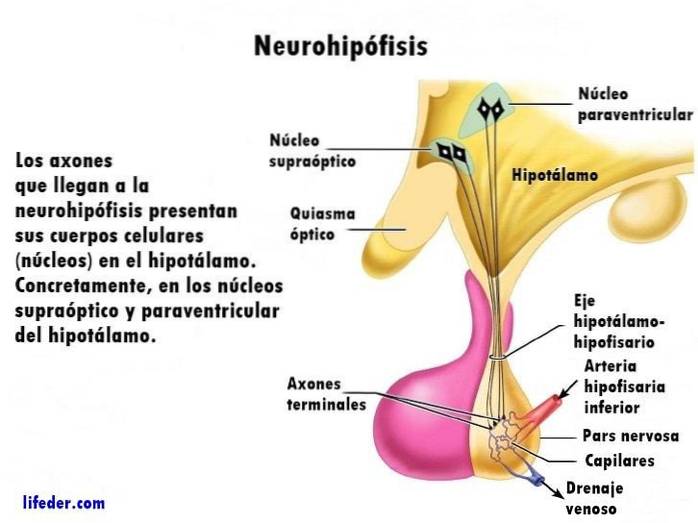
The axons (neuronal extensions) that reach the neurohypophysis present their cell bodies (nuclei) in the hypothalamus. Specifically, in the supraoptic and paraventricular nuclei of the hypothalamus.
These hypothalamic cell bodies create hormones that travel through the axons that cross the pituitary stalk, reaching the neurohypophysis. The latter can release hormones into the bloodstream directly.
To do this, the terminal buttons of the axons of the neurohypophysis connect with the blood capillaries. In these terminal buttons are stored the hormones that will be released into the blood when the body needs it..
It seems that the nerve impulses of the hypothalamus are those that control both the synthesis and the release of hormones accumulated in the neurohypophysis.
The neurohypophysis is formed by the differentiation of the neural ectoderm into the pars nervosa (or infundibular process), the infundibular stalk, and the median eminence.
The pars nervosa makes up most of the neurohypophysis, and is where oxytocin and vasopressin are stored. It has the unmyelinated axons of the neurosecretory neurons of the hypothalamus. In the hypothalamus are their cell bodies.
The pars nervosa is sometimes used synonymously with the neurohypophysis. However, this usage is incorrect.
Whereas, the infundibular stem or infundibulum is a structure that acts as a bridge between the hypothalamic and pituitary systems..
As for the median eminence, it is an area that connects with the pituitary stalk. There are authors who do not consider it part of the neurohypophysis, but of the hypothalamus.
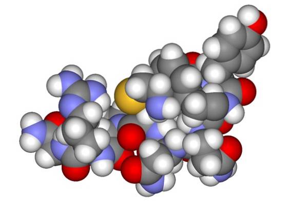
The hormones oxytocin and vasopressin are synthesized in the cell bodies of the hypothalamus. They then travel through the axons and accumulate in the terminal buttons, inside granules called Herring bodies..
Regarding the vasculature, the inferior pituitary arteries that come from the internal carotid artery are those that supply this structure. There is a network of capillaries that surround the axonal terminals, facilitating the released hormones to reach the blood..
The histological structure of the neurohypophysis is fibrous. This is due to the fact that it is constituted, above all, by unmyelinated axons of neurons of the hypothalamus. It has approximately 100,000 axons that carry hormones.
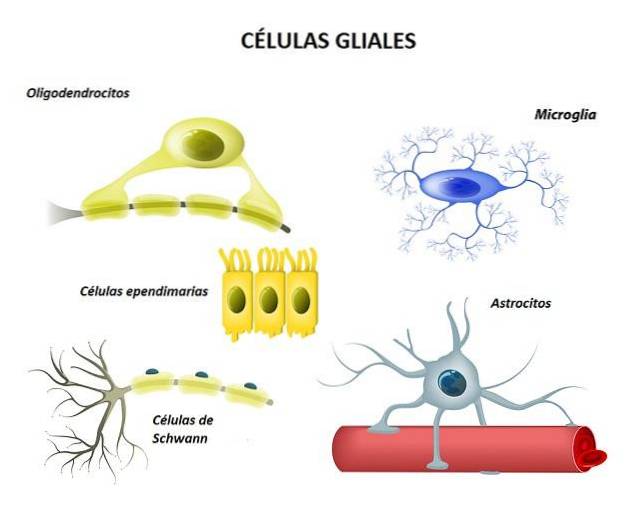
In addition, they also contain glial cells and a large number of capillaries. The latter are concentrated mainly in the ventral part, where there is a greater release of oxytocin and vasopressin into the blood. Many of the capillaries have small holes to facilitate the hormones reach the bloodstream.
An interesting and characteristic histological component of the neurohypophysis is the Herring bodies. They consist of widened protrusions located on the terminal buttons of the axons.
They have groups of neurosecretory granules, which contain oxytocin or vasopressin. They are usually linked to capillaries, and have an oval shape and a grainy texture.
On the other hand, specialized glial cells called "pituicytes" have been found in the neurohypophysis. Researchers believe that they could be actively involved in the regulation of hormone secretion. They have an irregular shape and an oval core.
The neurohypophysis stores and releases vasopressin and oxytocin. These hormones have effects associated with the autonomic nervous system.
Although the functions of oxytocin and vasopressin are different, their structure is very similar. Apparently, both come evolutionarily from the same molecule: vasotocin. This is still observed in some fish and amphibians.
The two hormones are synthesized in the nuclei (somas) of magnocellular neurons. Its name is due to its larger size and large soma. These are located in the supraoptic and paraventricular nuclei of the hypothalamus. Each neuron is specialized in the synthesis of only one type of hormone (either vasopressin or oxytocin).
For their synthesis, their precursors or prohormones are stored in neurosecretory vesicles that will process and convert them. In this process, enzymes convert their precursors, which are large proteins, into oxytocin and vasopressin.
On the other hand, the paraventricular and supraoptic nuclei of the hypothalamus secrete a substance called neurophysin. This consists of a protein that transports vasopressin and oxytocin through the hypothalamic-pituitary axis..
The following describes the hormones of the neurohypophysis:
Also known as antidiuretic hormone (ADH) for its effects on the kidney. Its main function is to regulate the secretion of water through urine..
Specifically, it stimulates fluid retention. In addition, it controls the vasoconstriction of peripheral blood vessels.
This substance contributes to the transport of milk during sucking, from the mammary glands to the nipples. In addition, it mediates the contraction of the smooth muscle of the uterus during orgasm. Like the contractions that occur at the time of delivery.
On the other hand, stress or emotional tension can alter the release of this hormone, even interfering with breastfeeding..
Interestingly, due to their similarity, these two hormones can cross-react. Thus, oxytocin at high levels has a mild antidiuretic function, while very high vasopressin can cause uterine contractions..
Tumors in the pituitary gland are relatively common. However, a tumor in the neurohypophysis is very rare. If present, it is usually accompanied by metastases and tumors in granule cells..
A congenital anomaly of the neurohypophysis called pituitary stalk disruption syndrome has also been found. Characterized by absent or ectopic neurohypophysis (developing in the wrong place), very thin or absent pituitary stalk, and anterior pituitary aplasia.
This results in deficiencies in the functioning of the pituitary gland, including the neurohypophysis. Some of the symptoms are hypoglycemia, micropenis, short stature, delayed development, low blood pressure and seizures.
Any damage or dysfunction of the neurohypophysis can cause problems in the secretion of vasopressin or oxytocin.
For example, in diabetes insipidus there is insufficient vasopressin release. In this disease, the body cannot concentrate urine. Those affected get to eliminate about 20 liters of diluted urine every day.
On the other hand, a very high vasopressin release causes the syndrome of inappropriate antidiuretic hormone secretion (ADH). This causes the body to retain more water than necessary, raising blood water levels too high..
Whereas, high doses of oxytocin can lead to hyponatremia. This means a very low concentration of sodium in the blood..


Yet No Comments