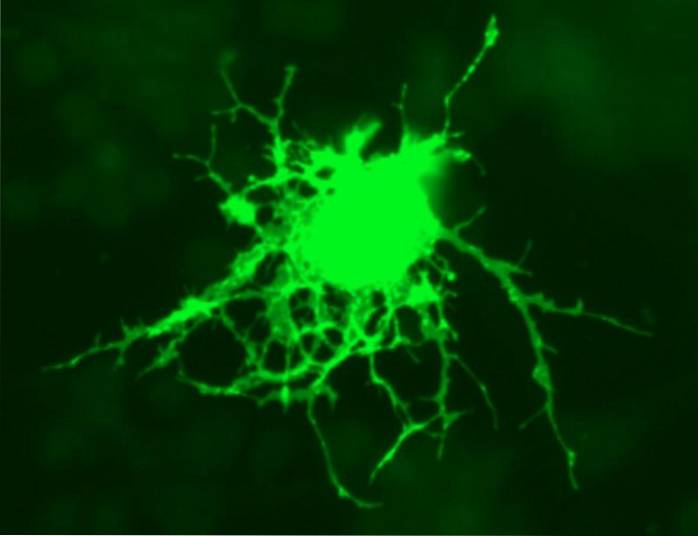
The oligodendrocytes or oligodendroglia are a specific type of macroglia cells. They are characterized by having a smaller size than that of astrocytes, as well as short and scarce extensions.
These types of brain cells primarily perform supportive and bonding activities. Likewise, they have the important function of generating the myelin sheath in the central nervous system..
At present, two main types of oligodendrocytes have been described in the neuroglia of the central nervous system: interfascicular oligodendrocytes, which are responsible for myelin production, and satellite oligodendrocytes, which appear to play roles in the sexual response..
This article reviews the main characteristics of oligodendrocytes. Their functions and their classification are discussed, and the formation process presented by this type of cells is explained..
Article index
Oligodendrocytes are a type of macroglial cell. That is, they are cells of the nervous tissue that are characterized by performing auxiliary functions, complementing the functioning of the main cells (neurons).
The term oligodendrocyte was introduced by the Spanish neurologist Pio del Rió Hortega and etymologically means little branched glia. In this sense, this type of cells are characterized by presenting short and fine branches, which can appear in the form of rows parallel to the nerve fibers.
There are currently two main types of oligodendrocytes: interfascicular oligodendrocytes and satellite oligodendrocytes..
The former are responsible for carrying out the myelination of the axons of the central nervous system. On the other hand, the latter have a much less documented functionality.
Regarding their formation, oligodendrocytes stand out for appearing late in development.
The development of oligodendrocytes is characterized by taking place in late stages. In fact, these types of cells originate when neurons have already been formed within the central nervous system..
Oligodendrocytes are formed from neurons that have migrated to their correct position, have been surrounded by glial cells, and have formed synaptic connections..
Specifically, oligodendrocytes arise from precursors that migrate through the white matter, from germinal areas of the ventricles and the central canal of the spinal cord..
Thus, the number of oligodendrocytes generated depends on the number of precursors that have migrated, divided, and differentiated. Likewise, programmed cell death in each brain region is also an important factor in the formation of this type of cells..
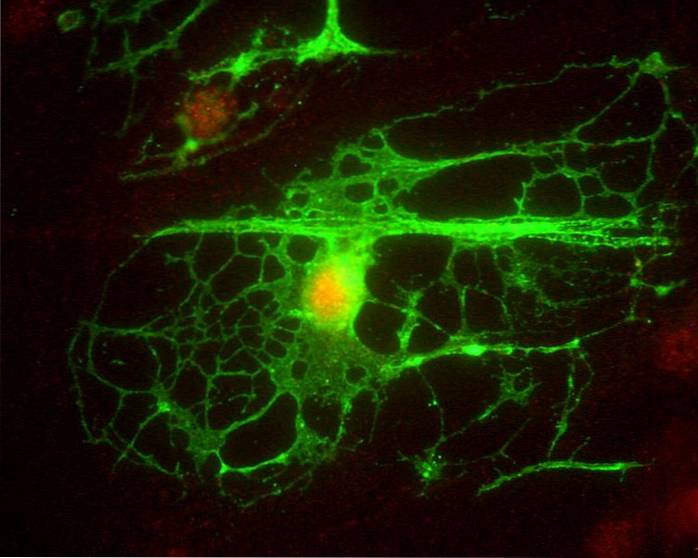
When the cells that have migrated differentiate, the precursors of the oligodendrocytes begin to generate processes that branch. This process generates a complex network and motivates a loss of migratory and proliferative capacity in the cell..
In contrast, the oligodendrocyte formation process causes the generation of the myelinating capacity of the cell, as well as the expression of specific components of milein.
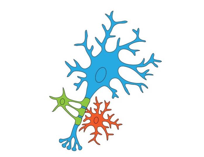
Myelin is a structure highly rich in lipoproteins that is responsible for wrapping the axons of the neurons of the nervous system. The adherence of this substance in neurons facilitates the electrical conduction of the nerve impulse and allows to increase the speed of synaptic transmissions..
The myelin sheath is generated by oligodendrocytes within the central nervous system while in the peripheral nervous system it is formed by Schwann cells..
Oligodendrocytes can be divided into two different types: interfascicular oligodendrocytes and satellite oligodendrocytes. The differentiation between these two types of cells rests mainly on their functionality, since they carry out different activities.
Interfascicular oligodendrocytes are responsible for the production of myelin and the isolation of the axon from neurons.
The satellite oligodendrocytes, on the other hand, present a certainly unknown activity. However, it is postulated that this type of cells could have an ejector function on the muscles of the cavernous tissue of the male sexual organ, thus participating in the sexual response and causing the sperm outflow process..
Anatomically, the two types of oligodendrocytes have similar characteristics. Both are characterized by containing few extensions. Likewise, its nuclei are rich in heterochromatin and its cytoplasms mainly contain ergastoplasma, free polyribosomes, a golgi apparatus and a high content of microtubules..
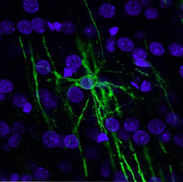
Oligodendrocytes are the cells of the central nervous system that are responsible for forming the myelin sheath of nerve fibers.
Thanks to the process of formation and maintenance of the myelin sheath, the axons of the central nervous system have an insulating coating that increases their speed of nerve conduction..
In addition, oligodendrocytes stand out for presenting extensions. Each of them allows the formation of different internodal myelin segments on the same axon or on different axons.
In fact, an oligodendrocyte can form up to 60 internodal segments, which is why this type of cells generate high amounts of myelin.
On the other hand, it should be noted that the myelin generated by oligodendrocytes presents a different formation process to that of Schwann cells in the peripheral nervous system..
Oligodendrocytes and their axons are not surrounded by a basement membrane. Thus, myelination begins around the sixteenth week of intrauterine life and continues during the postnatal period until most of the axons are myelinated..
Finally, satellite oligodendrocytes appear to play a role similar to that of the capsules of peripheral sensory ganglia. Certain studies postulate that this type of cells influence the biochemical environment of neurons and have been related to physiological processes related to sexual response.
The pathology that has been related to the functioning and activity of oligodendrocytes is multiple sclerosis.
This disease appears due to the loss of this type of cells and, therefore, of myelin sheaths on the axons of neurons.
In this sense, the loss of oligodendrocytes motivates the appearance of a series of symptoms that manifest the lack of myelin in neurons, such as loss of balance, muscle spasms, movement problems, coordination difficulties, tremor, weakness, constipation or alterations intestinal.


Yet No Comments