Craniometric points location and diameters of the skull
Craniometric points location and diameters of the skull
5360
376
Basil Manning
The craniometric points they are precise points located on the skull and on the face that serve as a reference for some linear and angular measurements. They are used for some anthropological studies and as anatomical landmarks for brain structures in neurosurgery and orthodontics..
They are grouped into those found in the frontal plane, in the superior plane, in the basal plane and in the lateral plane. Some points are single and others are bilateral or even.
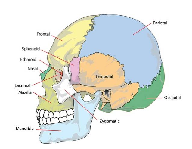
The skull and its bones (Image by Clker-Free-Vector-Images at www.pixabay.com)
Article index
1 Grouping
1.1 Frontal craniometric points
1.2 Craniometric points at the base of the skull
1.3 Upper craniometric points
1.4 Craniometric points of the lateral plane
2 Location
3 Diameters of the skull
3.1 Diameters of the face or the so-called viscerocranium
4 Images
5 References
Group
Frontal craniometric points
The frontal craniometric points are: the ophryo, the glabella, the nasion, the right and left dacrion, the right and left zigion, the rhinion, the cliff or nasospinal point, the prostion or alveolar point, the gnathion and the right and left gonion..
Craniometric points at the base of the skull
The craniometric points at the base of the skull are: the right and left zigion, the staphylion, the right and left portion, the basion, the opistion, the inion, and the opisthtocranion..
Upper craniometric points
The superior craniometric points are: the bregma, the right and left stepphanion, the vertex, the lambda, the obelion and the opisthtocranion (also observed at the base of the skull).
Lateral plane craniometric points
The craniometric points that are observed in the lateral plane are: the ophryo, the stepharion, the vertex, the opistocranion, the gabela, the nasion, the dacrion, the gnathion, the prostion, the nasospinal or cliff, the gonion, the pterion, the porion, the asterion and the inion.
Certain craniometric points can be defined and observed in various planes of the human skull, therefore some are repeated when defining those observed in each plane.
These reference points and the linear and angular measurements that derive from them change according to the typologies and allow anthropometric studies and facial reconstructions from skulls..
They are also used as references for some neurosurgical procedures by relating them to the underlying brain structures. Likewise, they are radiological reference points widely used in dentistry for the study of occlusion pathologies..
Location
There is a classification of craniometric points that does not make use of the planes of the skull, but rather groups the craniometric points into craniometric points of the neurocranium, sagittal and lateral, and the viscerocranium, sagittal and lateral.
Those of the sagittal neurocranium include bregma, vertex, lambda, opiscranion, inion, nasion, glabella, opistion, basion, sphenobasion, and hormone.
Bregma point (Source: NEUROtiker / CC BY-SA (http://creativecommons.org/licenses/by-sa/3.0/) via Wikimedia Commons)
Those of neurocraniumlateral They are the coronal, stepphanion, stenion, eurion, porion, mastoidal, pterion and asterion.
Points sagittal viscerocranial are the rhinion, nasospinal, subspinal, prostion, infradental, pogonium, gnathion, oral and staphylion points.
Points lateral viscerocranial include orbital, jugal, zinion, gonion, mental, temporal frontomalar, orbital frontomalar, zygomaxillary, lingual, koronion, medial kondylo, lateral kondylo.
The location of the main craniometric points is described below..
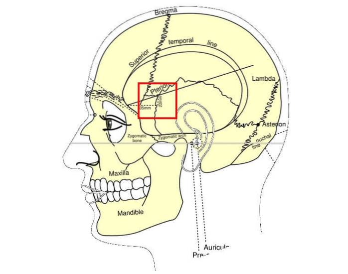
Pterion: the pterion is a point located in the center of the parietal-fronto-sphenoid suture. This suture is also called the pterytic suture, the anterior end of which is called the propterion and the posterior metapterion..
Asterion: the asterion is at the junction of the parietomastoid, lambdoid and occipitomastoid sutures.
Dacrion: the dacrion is at the junction of the frontal with the lacrimomaxilla.
Gonion: the gonion is located in the most distal and external part of the apex of the mandibular angle.
Nasion: the nasion is at the intersection or junction of the frontonasal suture with the internaal suture.
Eurion: the eurion is the point located in the most lateral prominent end of the skull, it can be located in the scale of the temporal bone or in the parietal bone. There is one right and one left.
Gabela: the gavel corresponds to the center of the frontal protrusion.
Gnathion: the gnathion is located in the midline of the jaw and is the lower point that corresponds to the lower part of the chin.
Zigion: the zigion is in the most protruding portion of the zygomatic arch.
Prosthion: the prostion is located in the maxillary bone between the alveolar processes of the upper incisors, which corresponds to the lowermost point of the anterior suture of the maxillary bone.
Inion: the inion corresponds to the most prominent point of the external occipital protuberance at the base of the skull.
Opistocranion: this craniometric point corresponds to the midpoint of the extreme posterior portion of the occipital bone.
Opistion: corresponds to the posterior or dorsal central point of the foramen magnum.
Basion: it is a point located in the most anterior or middle ventral portion of the edge of the foramen magnum.
Lambda: this point is located at the intersection site of the middle and lambdoid suture in the upper part of the skull in the posterior region.
Obelion: midpoint of an imaginary line that passes between the two parietal holes at the top of the skull.
Vertex: most prominent superior point of the sagittal suture in the superior plane of the skull.
Bregma: site of intersection or crossing between the coronal and sagittal sutures on the superior and anterior surface of the skull.
Skull diameters
By joining some craniometric points, the so-called diameters of the skull can be obtained, which, although they are widely used in anthropometry, are also used in dentistry through the radiographic identification of these points and diameters, especially used in orthodontics..
Maximum length of the skull: line joining the gavel and the opistocranion.
Length of the base of the skull: junction of the basion with the nasion.
Maximum width of the skull: virtual line that joins the two eurion points (one on each side)
Height of the skull: imaginary line joining the basion with the bregma
Combining the dimensions of these diameters, the cranial indices and their different categories are obtained. These are as follows:
Maximum width of the skull per 100 between the maximum length of the skull. The value of this relationship allows the following categories to be established:
Brachycephalic = 80.0 - 84.9
Dolichocephalus = 70.0 -74.9
Mesocranium = 75.0 - 79.9
Diameters of the face or the so-called viscerocranium
Length of the face: line that joins the basion with the prostion
Maximum width of the face: line that joins both the right and the left zigion
Total height of the face: line that joins the nasion point with the gnathion
Upper facial height: imaginary line that joins the nasion with the prostion.
The combination of any of these diameters allows to establish the facial indices with their respective categories.
The Total Facial Index or morphological index is equal to the total height of the face times 100 between the maximum width of the face. This index allows the following categories to be established:
Euriprosopo = 80.0 - 84.9
Mesoprosope = 85.0 - 89.9
Leptoprosopo = 90.0 - 94.9
The upper facial index is equal to the upper facial height times 100 times the maximum width of the face. The values of this index allow you to define the following categories:
Euriene = 45.0 - 49.9
Meseno = 50.0 - 54.9
Leptene = 55.0 - 59.9
Images
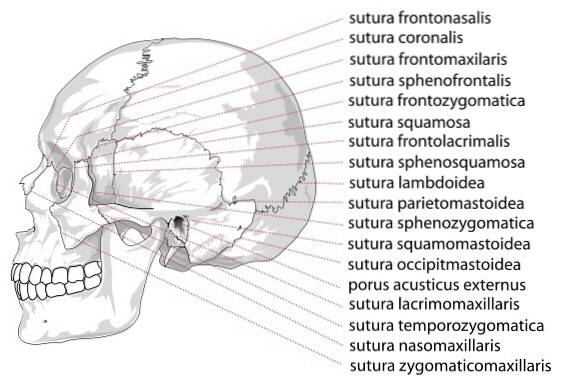
Pterion and other craniometric pointsHuman skull sutures
References
Cameron, J. (1930). Craniometric Memoirs: No. II. The Human and Comparative Anatomy of Cameron's Cranio-facial Axis. Journal of anatomy, 64(Pt 3), 324. Cameron, J. (1930). Craniometric Memoirs: No. II. The Human and Comparative Anatomy of Cameron's Cranio-facial Axis. Journal of anatomy, 64(Pt 3), 324.
de la Rúa Vaca, C. (1982). Dynamics of the craniometric points and the Klaatsch quadrilateral in the Basque Calvaria. Anthropology-Ethnography Notebooks, (1), 267-284.
Kendir, S., Acar, H. I., Comert, A., Ozdemir, M., Kahilogullari, G., Elhan, A., & Ugur, H. C. (2009). Window anatomy for neurosurgical approaches. Journal of neurosurgery, 111(2), 365-370.
Parzianello, L. C., Da Silveira, M. A. M., Furuie, S. S., & Palhares, F. A. B. (1996). Automatic detection of the craniometric points for craniofacial identification. Anais do IX SIBGRAPI'96, 189-196. Cotton, F., Rozzi, F. R., Vallee, B., Pachai, C., Hermier, M., Guihard-Costa, A. M., & Froment, J. C. (2005). Cranial sutures and craniometric points detected on MRI. Surgical and Radiologic Anatomy, 27(1), 64-70.
Ribas, G. C., Yasuda, A., Ribas, E. C., Nishikuni, K., & Rodrigues Jr, A. J. (2006). Surgical anatomy of microneurosurgical sulcal key points. Operative Neurosurgery, 59(suppl_4), ONS-177.
Toral Zamudio, T., Denis Rodríguez, P. B., & Jiménez Baltazar, C. A. (2019). Determination of tables of craniometric points based on cephalometry of Veracruz: study with recent corpses of medicolegal cases in the District of Xalapa, Ver. Mexican Journal of Forensic Medicine and Health Sciences, two(2), 1-10.


Yet No Comments