
The spermatogonia They are one of the different types of cells present in the testicles of animals. These are proliferative germ cells and are the only ones, in adult animals, capable of "self-renewal" and contribute to the formation of the next generation..
Many authors describe them as "the germ cells of males before meiosis" and, in animal species that present seminiferous tubules, these cells are found in the region corresponding to the basal lamina of said epithelium.
They are considered the “stem cells” of the male reproductive system, as they divide to maintain the number of cells in their population and to produce those cells that will differentiate into sperm.
Spermatogonia confer very special characteristics on male gonads, because thanks to their ability to divide, a male can produce an estimated 10 to 100 billion sperm throughout his life.
Article index
In all sexually reproducing animals, a small number of germ cells are formed during the early stages of embryonic development, destined solely for the production of the animal's sex cells (gametes).
Initially, these cells are indistinguishable in appearance between females and males, but this changes once these cells migrate and the gonadal tissue is formed, which, in males, is known as testis (s)..
The testes represent the only tissue class where meiosis occurs (just as the ovaries are for females). In them, the spermatogonia are the progenitor cells of the sperm, which are the differentiated sex cells, produced by meiosis and capable of fertilizing an egg.

Certain authors consider that the term "spermatogonia" can be used to refer to all cells in the testes that have not undergone meiosis.
Spermatogonia are generally round cells, characterized by a nucleus rich in chromatin (DNA + histone proteins). However, there are different types of spermatogonia, but their classification or nomenclature depends on the literature consulted..
Generally, many texts agree that spermatogonia divide by mitosis to form two types of cells, sometimes called A and B..
Type A spermatogonia are called replacement cells (undifferentiated cells), while type B spermatogonia are those that develop into spermatocytes, which then divide by meiosis.
Some authors, however, refer to these cells as part of three classes:
- The "mother" spermatogonia
- Proliferative spermatogonia
- Differentiated spermatogonia
The first two, that is, "stem cells" and proliferative spermatogonia, could be considered type "A", since they are responsible for the production of new spermatogonia and those spermatogonia that will later commit to differentiation..
The spermatogonia that will later differentiate into spermatocytes (equivalent to type "B", which will later become spermatozoa) undergo numerous mitotic divisions (this number may vary with the species), increasing the number of cells in the population of "B" spermatogonia..
The mitosis of these "differentiable" cells is, however, different from other types of mitosis, since cytokinesis is incomplete (the cells do not separate from each other after dividing in two), so all the resulting cells, called spermatocytes, held together as in a syncytium.
Type A spermatogonia are cells with very rounded nuclei that often, when stained with special dyes, are poorly colored. From the cytological point of view, many authors define two types of A spermatogonia, which are differentiated by their coloration in:
- Spermatogonia AD, from English dark, which means "dark"
- AP spermatogonia, from English pale, which means "pale"
Type B spermatogonia, on the other hand, are cells that are characterized by having nuclei with numerous nucleoli. Nucleoli are important intranuclear regions that are not bounded by a membrane but that perform very important functions, such as the synthesis of ribosomes..

These cells, when they have not started to differentiate, are not easily distinguishable from other spermatogonia, but they quickly begin to lengthen and undergo meiosis.
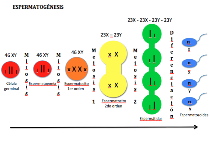
Spermatogenesis is defined as the process by which spermatogonia cells form spermatozoa and, at least in adult mammals, it is a process that occurs continuously until death.
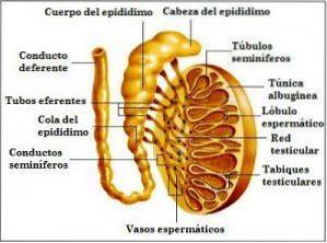
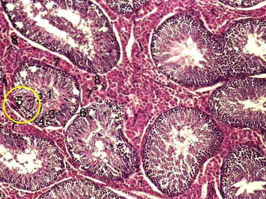
It occurs in the testicles, initially in structures called seminiferous tubules, which comprise about 90% of the testicular tissue. It has a mitotic and a meiotic phase.
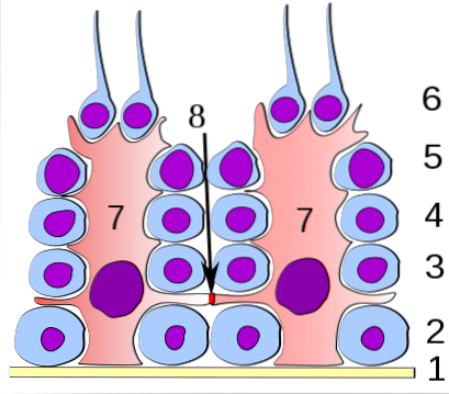
In the seminiferous tubules there are different types of cells, among them some called Sertoli cells are essential for nutrition and the support of the others.
These form a “hematotesticular” barrier that separates the intratubular epithelium in two:
- a basal compartment, where mitotic cells are exposed to the extratubular medium and
- a luminal compartment, where "postmeiotic" cells are exposed to an environment produced by Sertoli cells and germ cells
Spermatogonia are located in the basal compartment of the seminiferous tubules and are the cells that divide by mitosis to form new identical cells, some that remain as germ cells and others that differentiate.
As already mentioned, spermatogonia destined to differentiate into spermatozoa divide by mitosis, forming a kind of syncytium, since there is no complete cytokinesis (cell separation). It is these cells that subsequently divide by meiosis.
In general lines, a germinal spermatogonia can divide into two new cells or a pair of spermatogonia known as Apr, which remain united by an intercellular "bridge" (they do not complete cytokinesis).
These Apr cells can divide to form a chain of 4, 8, and occasionally 32 aligned A cells (Aal). All these cells are known as undifferentiated A spermatogonia or Aindif.
The aligned spermatogonia differentiate to become A1 spermatogonia. These cells divide successively (depending on the species), forming spermatogonia A2, A3, A4 and intermediate cells In, after which spermatogonia B are formed..
B cells divide to form primary spermatocytes which, upon completing different phases of meiosis, form secondary spermatocytes, from which haploid spermatids are formed.
Spermatids subsequently differentiate into sperm, the cells that then mature and whose main function is to fertilize the egg produced by a female of the same species..


Yet No Comments