
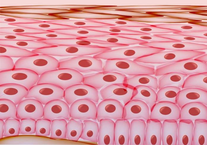
The keratinocytes They are the main group of cells that make up the epidermis, which is the thin layer located on the outermost part of the skin. They are named for the large number of keratin that they produce and contain inside, which makes them robust and resistant cells.
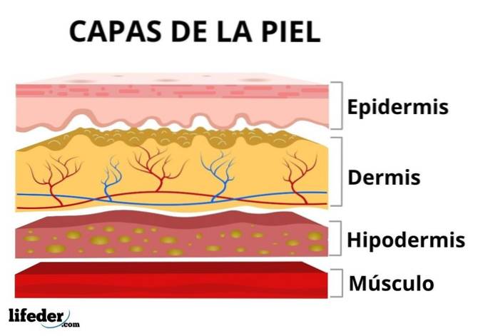
The skin is the largest organ in the human body, as it is the one that covers and protects its entire external surface. This fabric is made up of several layers, one on top of the other:
While the dermis and hypodermis are layers rich in blood vessels, nerves, glands, collagen, and hair follicles, the epidermis is rich in keratinocytes..
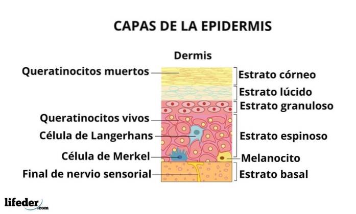
The epidermis is made up of three types of cells: keratinocytes, melanocytes, Langerhans cells and Merkel cells, the former being the most abundant.
It is between 0.007 and 0.12 millimeters thick (it is thickest on the soles of the feet and the palms of the hands) and is considered divided into 5 additional layers or regions: the basal stratum, the spinous stratum, the granular stratum, the stratum lucid and the stratum corneum, the most superficial.
Keratinocytes are cells that fulfill different functions, all closely related to each other, and that we can analyze according to whether they are protective, structural or immunological.
These cells are particularly specialized in protecting our body, since they form a resistant physical barrier that, to a large extent, prevents different substances or microorganisms from entering our system..
Another of its protective functions is the prevention of dehydration, since the epidermis formed by these cells also prevents, to some extent, spontaneous water losses.
They also participate in the prevention against heat loss under certain conditions.
Given that these cells represent more than 90% of all the cells present in the epidermis of our skin, one of the most obvious functions of these is to form this tissue, that is, to structure it..
Keratinocytes form strong associations with each other, generating an extremely orderly and resistant structure, where each cell is capable of always remaining in the same place..
Since these cells are literally "in the battle front", since they are found in the most superficial region of our body, it is necessary to point out that they fulfill important functions in wound healing, not only from the point of structural view, but also immunological.
When our skin suffers trauma, keratinocytes participate in a process known as epithelialization, where these cells migrate, multiply and differentiate to restore tissue, repairing the damage in question.
Epithelialization depends on a precise communication between the epidermis and the components of the immune system, which promotes these events by causing the release of different types of molecules (cytokines, chemokines, metalloproteinases, etc.) to the place where the wound has occurred..
Keratinocytes, as we have discussed, are found in the epidermis, which is the outermost layer of the skin of the human body, where they originate in the deepest region of said layer, known as basal stratum.
Once formed in the stratum basalis, these cells migrate to the surface, known as stratum corneum, where they differ, losing their nucleus and acquiring large amounts of keratin, the protein that characterizes them.
Keratinocytes change shape and appearance as they progress through their cell cycle and from one layer of the epidermis to the next. Thus, we can distinguish different types of keratinocytes depending on the stage we analyze:
They are the keratinocytes practically devoid of keratin and the only ones that retain the ability to divide, since they form the germinal basal layer, which gives rise to all the keratinocytes of the epidermis.
They are keratinocytes with the appearance of a spine or spike that are found, as their name may indicate, in the spinous layer, adjacent to the basal layer.
These cells are attached to each other through a special type of intercellular connection known as desmosoma which, seen under the microscope, has the appearance of a thorn.
They are the keratinocytes that advance towards the granular layer, which constitutes the hydrophobic tissue of our skin. They are cells that begin the production and storage of keratin and begin to lose the nucleus.
They are the keratinocytes that make up the stratum lucid and part of the stratum corneum. At this point in their life cycle they have lost the nucleus and have large keratin deposits, but they are dead cells. They are known as scales because of their appearance, since they are arranged one on top of the other and are elongated and flattened cells.
They form the stratum corneum, which is the outermost layer of the epidermis. Different authors define them as terminally differentiated keratinocytes and they are cells with very compact keratin stools, extremely flattened and that are shed from the surface of the body periodically.
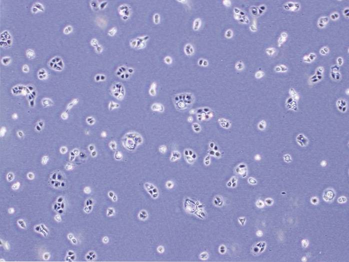
Human keratinocytes can be cultured under controlled conditions in vitro, in layers of a cell in thickness (monolayers) or in cultures with two-dimensional and three-dimensional structures, where they are provided with all the components they need to live.
The cells used for this purpose are generally derived from tissues such as the face, abdomen, chest and thighs of different donors..
Keratinocyte culture has different applications and utilities, as it is not only the basis of some epidermal studies related to the development and differentiation of the epidermis, but it is also exploited for drug effect studies and other pharmacological tests.
Likewise, the cosmetic and toxicological industries use these crops for the validation of the substances they commercialize; while they are also useful for research related to cancer, wound healing, tissue repair and other dermatological conditions, among others.


Yet No Comments