
The membrane receptors They are a type of cellular receptors that are located on the surface of the plasma membrane of cells, allowing them to detect chemical substances that by their nature cannot cross the membrane.
Generally, membrane receptors are integral membrane proteins specialized in the detection of chemical signals such as peptide hormones, neurotransmitters, and certain trophic factors; some drugs and toxins can also bind to these types of receptors.
They are classified according to the type of intracellular cascade to which they are coupled and which are the ones that determine the final effect on the corresponding cell, called the target cell or target cell..
Thus, three large groups have been described: those linked to ion channels, those linked to enzymes and those linked to protein G. The binding of ligands to receptors generates a conformational change in the receptor that triggers an intracellular signaling cascade in the target cell.
The signaling chains coupled to the membrane receptors make it possible to amplify the signals and generate transient or permanent responses or changes in the target cell. These intracellular signals are collectively called the "signal transduction system.".
Article index
The function of membrane receptors, and of other types of receptors in general, is to allow the communication of cells among themselves, in such a way that the different organs and systems of an organism work in a coordinated way to maintain homeostasis and respond to voluntary and automatic commands issued by the nervous system.
Thus, a chemical signal acting on the plasma membrane can trigger an amplified modification of various functions within the biochemical machinery of a cell and trigger a multiplicity of specific responses.
Through the signal amplification system, a single stimulus (ligand) is capable of generating immediate and indirect transient changes and long-term changes, modifying the expression of some genes within the target cell, for example.
Cellular receptors are classified, according to their location, into: membrane receptors (those that are exposed in the cell membrane) and intracellular receptors (which can be cytoplasmic or nuclear).
Membrane receptors are of three types:
- Bound to ion channels
- Linked to enzymes
- G protein-linked
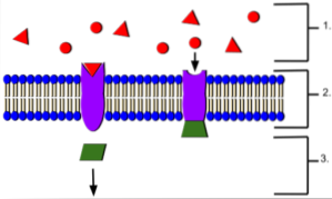
Also called ligand-gated ion channels, they are membrane proteins composed of between 4 and 6 subunits that are assembled in such a way that they leave a central channel or pore, through which the ions pass from one side of the membrane to the other..
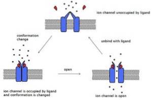
These channels cross the membrane and have an extracellular end, where the binding site for the ligand is located, and another intracellular end that, in some channels, has a gate mechanism. Certain channels have an intracellular ligand site.
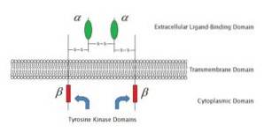
These receptors are also transmembrane proteins. They have an extracellular end that presents the binding site for the ligand and that have an enzyme associated with their intracellular end that is activated by binding the ligand to the receptor.
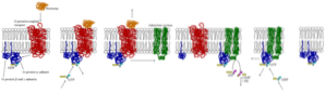
G-protein-coupled receptors have an indirect mechanism for the regulation of the intracellular functions of target cells that involves transducer molecules called GTP-binding or binding proteins or G-proteins..
All of these G-protein-linked receptors are made up of a membrane protein that crosses the membrane seven times and are called metabotropic receptors. Hundreds of receptors linked to different G proteins have been identified.
In ion channel-bound receptors, the binding of the ligand to the receptor generates a conformational change in the structure of the receptor that can modify a gate, move the walls of the channel closer or further apart. This modifies the passage of ions from one side of the membrane to the other..
Receptors bound to ion channels are, for the most part, specific for one type of ion, which is why receptors for K +, Cl-, Na +, Ca ++ channels, etc. have been described. There are also channels that let two or more types of ions pass.
Most enzyme-linked receptors associate with protein kinases, especially the enzyme tyrosine kinase. These kinases are activated when the ligand binds to the receptor at its extracellular binding site. Kinases phosphorylate specific proteins in the target cell, modifying its function.
G protein-linked receptors activate cascades of biochemical reactions that end up modifying the function of various proteins in the target cell..
There are two types of G proteins which are the heterotrimeric G proteins and the monomeric G proteins. Both are bound to GDP inactively, but by binding the ligand to the receptor, GDP is replaced by GTP and the G protein is activated..
In heterotrimeric G proteins, the GTP-bound α subunit dissociates from the ßγ complex, leaving the G protein activated. Both the α subunit bound to GTP and the free ßγ can mediate the response.
Monomeric G proteins or small G proteins are also called Ras proteins because they have been described for the first time in a virus that produces sarcomatous tumors in rats..
When activated, they stimulate mechanisms mainly related to vesicular traffic and cytoskeletal functions (modification, remodeling, transport, etc.).
The acetylcholine receptor, linked to a sodium channel that opens upon binding to acetylcholine and causes depolarization of the target cell, is a good example of membrane receptors linked to ion channels. In addition, there are three types of glutamate receptors that are ionotropic receptors..
Glutamate is one of the most important excitatory neurotransmitters in the nervous system. Its three types of ionotropic receptors are: NMDA (N-methyl-D-aspartate) receptors, AMPA (α-amino-3-hydroxy-5-methyl-4-isoxazole-propionate) and kainate (acid kainic).
Their names are derived from the agonists that activate them and these three types of channels are examples of non-selective excitatory channels, since they allow the passage of sodium and potassium and in some cases small amounts of calcium..
Examples of enzyme-linked receptors are the insulin receptor, the family of TrK receptors or neurotrophin receptors, and the receptors for some growth factors..
Major G-protein-coupled receptors include muscarinic acetylcholine receptors, β-adrenergic receptors, olfactory system receptors, metabotropic glutamate receptors, receptors for many peptide hormones, and rhodopsin receptors of the retinal system..


Yet No Comments