
The osteotendinous reflex or myotatic, Also known as a deep reflex or muscle stretch reflex, it is an involuntary motor response to an external stimulus, characterized by the contraction of the muscle that opposes a provoked stretch.
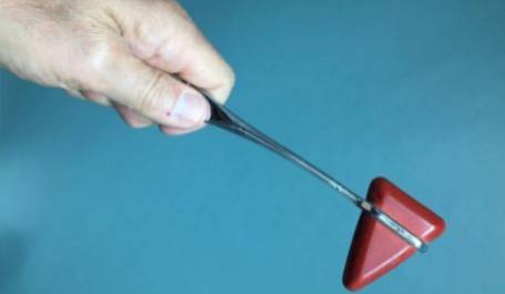
This reflex is intentionally generated during clinical evaluation when the clinician, using a small hammer, gently strikes a muscle tendon, causing it to contract. There are many examples of tendon reflexes; among the most popular is the knee-jerk reflex.
The response of this reflex to the stimulus in the knee is the contraction of the quadriceps femoris and the involuntary “kick”. The biceps reflex also stands out, in which the biceps brachii tendon is stimulated in the elbow crease and the arm is contracted; the answer resembles the vulgar gesture known as "sleeves cut".
Other reflexes belonging to this group are tricipital, styloradial, ulnar pronator, aquilane, mediopubian, nasopalpebral, supraciliary and masseter, among others..
Article index
Like any spinal reflex mechanism, the osteotendinous or myotatic reflex consists of: receptor, afferent pathways, nervous center and efferent pathways.
The receptor that is activated in this pathway is called the muscle spindle. Each receptor is made up of a few muscle fibers surrounded by connective tissue.
These fibers are called intrafusal fibers, in order to differentiate them from the other fibers that make up the muscle and which are called extrafusal fibers..
In turn, intrafusal fibers are of two types: nuclear sac fibers and nuclear chain fibers. In the nuclear sac fibers there are primary nerve endings from which the rapidly conducting afferent fibers originate..
The primary endings and fast conducting fibers are those that participate directly in the reflex through their connection with motor neurons..
The impulse travels through the axons of the sensory neurons of the muscle and reaches the posterior horn of the spinal cord..
It is found in the spinal cord and is made up of a sensory neuron and a motor neuron..
They are formed by the axons of motor neurons.
The most characteristic of the osteotendinous reflex is its monosynaptic condition, which implies that only one synapse is made between the afferent and efferent neurons..
The receptor senses the stretch, which stimulates the nerve fiber inside the muscle. The nerve impulse thus generated circulates along the sensory nerve, penetrating the spinal cord through the posterior roots..
Then it synapses with the neuron of the anterior root destined for the previously stretched muscle, where the response that travels through the efferent pathway is generated. The circuit is closed with the contraction of said muscle.
It is a simplified summary of the tendon reflex, because other more complex elements may be present.
A more complete explanation includes the intramedullary circuits of association that inhibit the antagonist or opposing musculature, and the superior structures that modulate this reflex arc..
In addition, the pyramidal and extrapyramidal bundles influence the reflex with an inhibitory action by the former and excitatory by the latter..
Like most proprioceptive, myotatic or stretching reflexes, the osteotendinous reflexes have protective functions against excessive stretching, they serve as the basis of muscle tone and, in addition, with their clinical evaluation they allow to assess the integrity of the nerve segments that are involved in the same.
To properly interpret stretch reflexes, the following should be taken into account:
- Stretch reflexes are sought by provoking the short, abrupt stretch when the tendon is struck with a reflex hammer. The hammer blow should be strong enough to elicit the stimulus, but not so strong as to cause pain to the examined patient..
- It is preferable to use rubber hammers.
- The evaluation should always be done on both sides of the body when it comes to a "mirror" muscle..
- To obtain a better response, it is convenient that the patient is relaxed; the muscle to be explored should also be in a maximally short or distended position.
Although numerous stretch reflexes are known, it is sufficient for the physician to know and explore the following:
The patient should have a half-open mouth. The examiner places a thumb on the examinee's chin and strikes it with the hammer. The answer is a contraction of the masseters and temporal bones, which leads to the closure of the mouth..
The patient flexes the forearm at a right angle at the elbow. The examiner places the forefinger or thumb on the biceps brachii tendon and strikes the hammer on his own finger. The answer is the flexion of the forearm with slight supination on the forearm.
The patient flexes the forearm at a 120º angle with the arm. The hammer is struck directly on the muscle tendon at the level of its insertion at the elbow. The answer is the extension of the forearm over the arm.
The patient flexes the forearm at a right angle and semi-pronation. Percuss the styloid process of the radius. The answer is flexion and supination of the forearm.
The patient should be seated with the legs pendulous or crossed. It is struck on the quadriceps tendon below the patella. The answer consists of the extension of the leg on the thigh.
The patient lies on his stomach, the knee of the lower limb to be explored is flexed and the foot in dorsal semi-flexion. The Achilles tendon is struck near its insertion on the calcaneus, in the vicinity of the ankle. The answer is a slight plantar flexion of the foot..
A reflex can show damage or illness due to lack or excess of response. In the first case, we can speak of hyporeflexia, when the response is diminished; or areflexia, when there is no response at all.
The excess response is known as hyperreflexia. It will be up to the doctor to determine the causes of these altered responses, make the diagnosis and establish treatments.


Yet No Comments