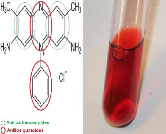
The safranin It is a meriquinoid dye, named for having in its chemical structure 2 benzenoid rings and 2 quinoid rings, the latter being the ones that provide the red color.
It is also called dimethyl safranin or basic red 2 in its short form, since its scientific name is 3,7-diamino-2,8-dimethyl-5-phenyl-phenaziniumchloro dimethyl safranin and the chemical formula is CtwentyH19N4 Cl.
There is a variant called trimethyl-safranin but there is no significant difference between the two substances.
Safranin is a monochromatic dye and, depending on the characteristics of the chemical formula, it is a positively charged substance. Therefore, it has an affinity for negatively charged structures. These structures will be stained red.
This property gives it applicability in many histological techniques to stain various cellular structures, both in eukaryotic and prokaryotic organisms..
Safranin is used as a contrast dye in important and well-known techniques for routine use in bacteriology. These techniques are: the Gram-Hucker stain, the Schaeffer Fulton stain for spores or the staining of bacterial capsules, among others..
Article index
The color of saffron (a spice obtained from the stigmas of the flower of Crocus sativus) was the inspiration to name this coloring. From the term saffron comes the name of safranin. This is due to the great similarity between the color of saffron and the coloring that this dye provides..
Safranin is obtained as crystals or in powder, both presentations being soluble in water. The safranin dye is odorless. Stains structures red. The structures that attract safranin dye are called safranophiles..
Structurally safranin is complex, it has two benzenoid rings at the ends and in the center are located the two quinoid rings where the N cation is found.+. The center of the structure is the system in charge of providing the color. Due to this characteristic, this colorant is classified within category II..
Safranin is used to stain various structures. Especially highlights the Kulchitsky cells present in the gastrointestinal tract, also called enterochromaffin cells.
It is capable of staining microorganisms belonging to the family Rickettsiaceae. Likewise, it is used in various techniques, such as the Koster method, a modified used for the staining of bacteria of the genus Brucella.
On the other hand, safranin is used in the Schaeffer Fulton spore staining technique and in the Gram-Hucker staining. In both techniques, safranin works as a contrast dye.
In the first, the spores take the color of malachite green and the rest of the structures are red by safranin. In the second, the Gram negative bacteria lose the color of the violet crystal in the discoloration step, therefore safranin is the one who stains the Gram negative bacteria red.
Additionally, safranin is used in bacteriology to prepare Brucella agar media with a 1: 5000 dilution of safranin. This medium serves to differentiate the species Brucella suis of the rest of the species. Brucella melitensis Y Brucella abortus they grow in this environment but B. suis is inhibited.
In the agro-industrial field, safranin has been used at 2.25% and diluted 1:10 to stain samples of the stem of the sugarcane plant.
This plant is commonly affected by the bacteria Leifsonia xyli subsp. xyli, who damages the xylem of the plant. The stained stems are evaluated to determine the function of the xylem vessels..
A blood or tissue smear is placed in a buffer solution (phosphate buffer pH 7.6). It is allowed to dry spontaneously and then covered with methylene blue for 3 minutes and counterstained with safranin. Rickettsiae are colored blue, contrasting with the red background.
A smear is made and flamed in the lighter for fixation. Subsequently, it is covered with a mixture of 2 parts of aqueous safranin saturated with 3 parts of 1 mol / L KOH solution, for 1 minute. It is washed with distilled water and counterstained with 1% carbolic methylene blue..
If the sample contains bacteria of the genus Brucella these will appear orange on a blue background.
A mixture of bacterial suspension is made with India ink and safranin is added to it. Under the microscope, a reddish halo will be observed around each bacterial capsule with a black background..
A smear is made with the bacterial suspension. Then it is fixed to heat. It is covered with 5% malachite green, flaming frequently until fumes are emitted. The process is repeated for 6-10 minutes. Finally, it is washed with water and counterstained with 0.5% safranin for 30 seconds. Bacilli stain red and spores green.
A smear is made with bacterial suspension and fixed in the heat. Cover the slide with crystal violet for 1 minute. Lugol is then placed as a mordant solution for 1 minute. Subsequently, it is bleached with acetone alcohol and finally counterstained with safranin for 30 seconds..
Gram positive bacteria stain bluish purple and Gram negative bacteria red.
Some laboratories have stopped using the Gram-Hucker technique to adopt the modified Gram-Kopeloff technique. In the latter, safranin is replaced by basic fuchsin. This is because safranin weakly stains species of the genera Legionella, Campylobacter Y Brucella.
Tissue sections from the gastrointestinal tract are stained with silver chloride. It is then decolorized with sodium thiosulfate and finally counterstained with safranin.
Kulchitsky cells are distinguished by the presence of blackish-brown granules..
Because safranin has a positive charge, it binds very well to the carboxyl and sulfate groups of glycosaminoglycans. These are part of the proteoglycans that make up the articular cartilage. In this sense, when staining with safranin O, it is possible to identify whether or not there is loss of cartilage..
The loss of cartilage tissue can be measured through the Mankin scale or also called the osteoarthritis scale..
The technique is explained below: the histological section is immersed in a tray with Weigert's iron hematoxylin solution, then passed through acid alcohol and washed with water.
Continue the coloring process by immersing the sheet in fast green, it is washed with acetic acid and now it is immersed in safranin O. To finish the process, it is dehydrated using alcohols at different concentrations in ascending order. The last step requires xylene or xylol for the sample to clarify.
The slides are conditioned with Canada balsam or similar to be observed under a microscope..
With this technique, the nuclei are stained black, the bone green and the cartilage where the proteoglycans are found red.
Pérez et al in 2003 proposed a simple and inexpensive technique to dye macroalgae. The samples are prepared in paraffin histological sections. The sections are fixed with 1% glycerin, allowing them to dry completely. It is then placed in xylol to remove the paraffin.
The section is rehydrated by passing it through a series of trays containing ethanol in different degrees of concentration (descending order), for 2 min in each one..
Subsequently, it is stained for 5 minutes with a 3: 1 mixture of 1% safranin with 1% toluidine blue, both prepared with 50% ethanol. Three drops of picric acid are added to the mixture, which acts as a mordant..
Then it is dehydrated by going through the alcohol trays again, but this time in an ascending way. Finally, it is rinsed with xylol and the sample is prepared with Canada balsam to be observed..
Fortunately, safranin is a dye that does not represent a danger to those who handle it. It is a harmless colorant, it is not carcinogenic and it is not flammable.
Direct contact with the skin or mucous membranes can cause a slight redness in the area, without major complications. For this, it is recommended to wash the affected area with plenty of water.


Yet No Comments