
The Giordano's sign It is the painful response of an individual to the stimulus caused by the doctor when he taps his hand gently on the sides of the lower back, on the lumbar spine. It is evidenced in patients with upper urinary tract infections or pyelonephritis.
Kidney infections have very dramatic symptoms. This means that the patient looks really sick with a very high fever, vomiting, and malaise. Low back pain is a common feature. There may also be pain in the groin and towards the genitals. However, these symptoms are not specific to kidney disease..
The pain can be moderate to severe, depending on the patient's pain threshold, which is the ability of each person to withstand pain. But when the sensitivity of the lumbar area is present without the need for aggressive maneuvers, the doctor can make the diagnosis and begin an appropriate treatment plan.
When the doctor relates the questioning of the patient with the laboratory tests, the Giordano sign becomes very specific for the diagnosis of pyelonephritis..
Article index
Giordano's sign is a physical examination maneuver that consists of percussion with the edge of the hand, at the level of the lumbar region. If the patient has pain, Giordano's sign is positive and indicates that the patient has kidney disease.
This maneuver was described by the doctor Davide Giordano (1864-1954), who enriched the surgical field with his important contributions in the specialties of gynecology, abdominal surgery, urology and even traumatology.
It differs from other semiological maneuvers for renal exploration since in this case, the percussion is performed with the edge of the hand. In other maneuvers, such as Murphy's, a lumbar percussion is also performed, but with a closed fist.
Also known as Pasternacki's sign, it is a clinical sign that, together with the medical history and laboratory results, is quite specific for kidney disease.
To achieve evidence of pain, it is not necessary to exert a great force when performing the maneuver since with a minimal blow the patient will present pain.
The pain occurs due to inflammation of the kidney parenchyma due to infection or the presence of stones or stones in the ureters. For this reason, with a minimal rebound caused by manual percussion of the lumbar area, the patient presents a pain of great intensity.
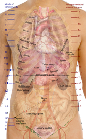
The kidney is a paired organ, there is a right and a left one, which is part of the upper urinary system. It is located in the abdomen behind the peritoneal lamina, which is the membrane that covers most of the abdominal organs..
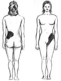
Each kidney is on one side of the lumbar spine and topographically they are located towards the area where the ribs make an angle with the spine. This area is known as the costovertebral angle..
The most common diseases of the kidneys are kidney stones or lithiasis and infection by bacteria or acute pyelonephritis.
Since the function of the kidneys is the filtering of toxic products from the blood, any disease that affects its filtering capacity can have serious consequences on the health of the patient. That is why the diagnosis must be timely in order to initiate adequate and timely treatment..
Acute pyelonephritis is a disease of the upper urinary tract. The upper urinary tract is made up of the kidneys and ureters, which are the outflow tubes that connect the kidneys to the bladder..
The flow of urine is downward. Urine is formed in the kidneys, from there it passes to the ureters to be stored in the bladder until the moment of its expulsion through urination, through the urethra.
Urinary infections are most often found in female patients. This is because the length of the urethra is shorter in women than in men, which allows bacteria to infect more easily..

Other common causes of pyelonephritis is the so-called reflux vesicoureteral, this is that there is an inverted flow of urine and as it descends towards the bladder, it also begins to rise from the bladder towards the ureter finally reaching the kidney and stagnating there.
Stagnant urine in the kidney is contaminated with bacteria that end up infecting the kidney surface.
Stones or stones in the kidneys is a disease known as renal lithiasis. When the stones are large enough, they can obstruct the lumen of the ureters, making urine unable to flow normally into the bladder..
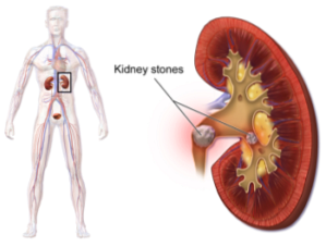
The urine that remains between the ureter and the kidney ends up being contaminated and infecting the kidney parenchyma.
Patients with immunosuppressive conditions, low defenses, are more prone to pyelonephritis. Thus, patients with poorly controlled diabetes, HIV, lupus erythematosus, among other diseases, have a higher risk than the rest of the population of having high urinary tract infections..
In these cases, the infection can present with different symptoms that make it difficult to diagnose and can become infected with bacteria that are difficult to treat..
The diagnosis of pyelonephritis is made from the questioning of the patient, laboratory tests and especially the physical examination.
The patient presents with discomfort when urinating, pain, burning or difficulty in urination. This discomfort increases with the passing of the hours and can even lead to urinary incontinence.
Pain in the lumbar region is also one of the symptoms that are frequently found in patients with this type of disease..

The laboratory tests that are ordered are blood tests, which can indicate infection and the simple urine test, which will clearly reveal the typical signs of a urinary infection which are cloudy urine with lots of bacteria and, in some cases, blood and other cells.
Regarding the physical examination, the most common is that the doctor finds a patient with a high fever (greater than or equal to 39 ° C), general malaise and pain in the lower back..
It is at that moment when maneuvers are performed to locate the pain towards the kidney. One of the most reliable maneuvers is the one described by Giordano to show pain in the location of the affected kidney.
The treatment of kidney infection depends on the type of bacteria that is contaminating the kidney, the cause of the infection and the underlying pathologies of each patient..
The main thing is to do a urine culture, which is a special test that isolates the specific bacteria and shows which antibiotic attacks it most effectively. By having this result, an appropriate treatment can be started.
If the patient also has some other medical condition, such as diabetes, that disease should also be treated as it aggravates the infection.
If pyelonephritis occurs due to kidney stones or stones, once the infection improves, the specialist must clean the kidney of them, either through medical or surgical treatment..


Yet No Comments