
Psoas sign what is it, psoas muscle anatomy

The psoas sign it is a clinical response associated with irritation of the peritoneum, which is the layer that lines the abdominal cavity. This sign becomes evident when the doctor performs the psoas maneuver for abdominal pain.
The maneuver consists of asking the patient to stretch his right leg back while lying on the left side. The sign is positive if the patient has pain when performing the movement. The maneuver manages to activate the psoas, which is a large muscle found in the abdominal cavity and which has important functions in gait and stability..
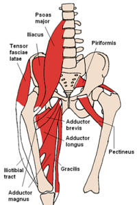
Being within the abdominal cavity, the psoas muscle is in contact with the peritoneal layer. This contact achieves that, when the peritoneum is inflamed by an infectious process of the abdomen, the active movement of the psoas highlights the pain.
This sign is considered one of the main ones to take into account when it is suspected that the patient is going through a process of inflammation of the cecal appendix, especially when this organ is in a posterior position close to the muscle..
Although the psoas sign is indicative of any infectious process that causes inflammation of the peritoneum, it is more frequently associated with acute appendicitis. The sign has been described by several surgeons throughout history without attributing its description to any one in particular..
Article index
- 1 Anatomy: psoas muscle
- 1.1 Origin
- 1.2 Function
- 1.3 Anatomical relationships
- 2 What is the sign of the psoas?
- 2.1 Clinical considerations
- 3 References
Anatomy: psoas muscle
The psoas is a muscle that is located inside the abdomen behind the peritoneal layer. It is one of the largest and most important retro-peritoneal organs.
Made up of two fascicles called psoas major and psoas minor, it is one of the most important muscles for stability and gait..
Source
The tendons of origin of the psoas attach to the last dorsal and first lumbar vertebrae.
The longest bundle of the psoas, called the psoas major, originates from the last thoracic or dorsal vertebrae and the first four lumbar vertebrae. It is made up of two segments, one superficial and the other deep..
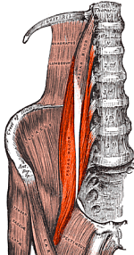
The deep segment is the one that originates from the first four lumbar vertebrae (L1-L4), while the superficial segment originates from towards the outer edge of the last dorsal vertebra (T12) creating a firm tendinous structure by joining with the adjacent ligaments. to the vertebral discs.
These two segments unite to form the muscular body of the psoas, which in its lower portion joins with the iliac muscle, giving rise to the muscle known as iliopsoas..
The smallest bundle of the psoas, called the psoas minor, is a thin segment of the psoas that originates from the last dorsal and first lumbar vertebrae (T12-L1). It is a long portion that reaches the pubis and its function is to support the psoas major.
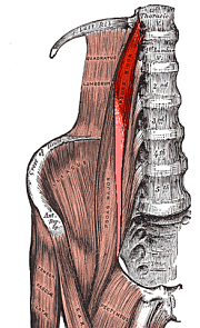
The psoas minor has many anatomical variations and is considered an inconstant muscle since it is absent in 60% of individuals.
Function
The psoas performs important functions in gait and balance. Its tendinous attachments, which go from the thoracic spine to the femur, connect the trunk with the lower limbs..
The activation of the psoas achieves the flexion of the hip, the maintenance of the upright position and, in conjunction with other muscles, the incorporation from the horizontal to the vertical position (lying down to standing).
Anatomical relationships
The psoas is a retro-peritoneal muscle, this means that it is not covered by the sheet called the peritoneum that covers the abdominal organs..
Its long history makes it related to several intra-abdominal structures including the kidneys and the colon..
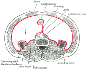
On the right side, the colon is in a more posterior position, and in some anatomical variations, the cecal appendix is located even more posteriorly, coming into contact with the psoas..
When there is an infection in the abdomen, the peritoneum reacts by triggering an inflammatory process that in a few hours installs a picture of abdominal pain.
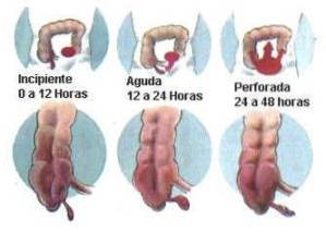
The proximity of the cecal appendix to the psoas muscle causes an irritation of the fibrous layer that covers the muscle, which is why it becomes inflamed, triggering pain with its movement.
What is the sign of the psoas?
To highlight the sign of the psoas, the doctor must perform the maneuver of forced active movement of the muscle, this means that the patient himself must perform a movement, without help, and force the limb as much as possible in the direction that it is asks you.
The patient should be lying on the left side. Once in that position, he is asked to straighten his right leg and perform a forced movement (as much as possible) of extension backwards. The sign is positive if this movement causes the patient such pain that the movement must be interrupted.
Another way to achieve a positive psoas sign is with the patient lying on their back. In this position you are asked to raise your leg about 50 cm off the bed. The doctor places his hand on the patient's thigh and exerts downward pressure requesting the patient to try to overcome this force by raising the leg further..
The sign is considered positive if pain of such magnitude is triggered that the patient must interrupt movement.
In both cases, what is sought is the activation of the muscle so that it causes the inflamed peritoneal lamina to rebound and cause pain.
Clinical considerations
The positive psoas sign is indicative of an abdominal inflammatory process. It can be specific for acute appendicitis when it is evaluated in conjunction with other clinical signs and when it is related to the evolution of the pain that the patient presents..
The appendix presents anatomical variations in a significant percentage of people. One of the most common is the appendix located behind the cecum, called the retrocecal appendix..
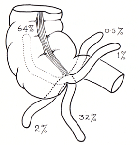
In the retrocecal position, the appendix is in direct contact with the psoas muscle separated only by the thin peritoneal lamina.
Acute appendicitis is an infectious condition that causes a significant peritoneal inflammatory process. This process takes 4 to 6 hours to install..
During this time and with the passing of the hours, the movements that the peritoneum rebound causes great pain in the affected individual..
The inflammation that triggers the peritoneum also manages to irritate and inflame nearby organs. In this way, the psoas sign causes pain through two mechanisms.
At the moment of activating the muscle, and more so if it is forced, the inflamed peritoneum layer has the rebound movement that is required to bring out the pain. In addition, the body of the muscle begins to swell due to the proximity of the infected organ, so the muscle activation movements cause pain.
The psoas sign alone does not establish a diagnosis, but when evaluated together with the rest of the clinical signs, examinations and symptoms of the patient, it can guide towards the different pathologies that cause peritoneal irritation..
References
- Sherman R. (1990). Abdominal Pain. Clinical Methods: The History, Physical, and Laboratory Examinations. 3rd edition, chapter 86. Boston. Taken from: ncbi.nlm.nih.gov
- Rastogi, V; Singh, D; Tekiner, H; Ye, F., Mazza, J. J; Yale, S. H. (2019). Abdominal Physical Signs and Medical Eponyms: Part II. Physical Examination of Palpation, 1907-1926. Clinical medicine and research. Taken from: ncbi.nlm.nih.gov
- Sajko, S; Stuber, K. (2009). Psoas Major: a case report and review of its anatomy, biomechanics, and clinical implications. The Journal of the Canadian Chiropractic Association. Taken from: ncbi.nlm.nih.gov
- Siccardi MA, Valle C. (2018). Anatomy, Bony Pelvis and Lower Limb, Psoas Major. StatPearls. Treasure Island (FL). Taken from: ncbi.nlm.nih.gov
- Mealie, CA; Manthey, DE. (2019). Abdominal Exam. StatPearls. Treasure Island (FL). Taken from: ncbi.nlm.nih.gov
- Jones, MW; Zulfiqar, H; Deppen JG. (2019). Appendicitis. StatPearls. Treasure Island (FL). Taken from: ncbi.nlm.nih.gov


Yet No Comments