
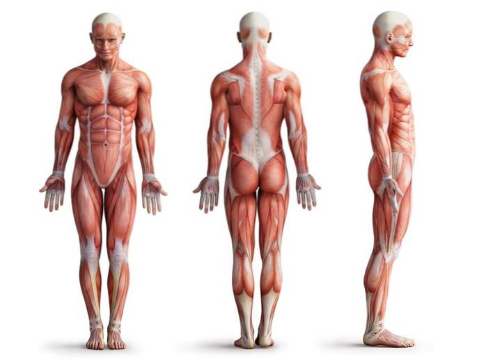
The muscular system It is the system of the human body that is made up of all the muscles. It includes both the muscles associated with the skeleton, which allow the body to move, and the muscles that make up the heart and internal visceral organs..
Muscles comprise a very special tissue of our body, since it has the ability to contract and, with it, produce movements. These movements are generally the result of the action of the nervous system on the groups of cells that form muscle mass..
Like other body systems, the muscular system allows us, in one way or another, to interact with the environment that surrounds us, responding to its changing conditions and to our own physiological needs: hunger, thirst, reproduction, shelter , flight or defense, among others.
Our body has many different muscles, many of which are specialized in very specific functions..
The digestive, circulatory and respiratory systems are good examples of the participation of the muscular system in functions other than those of movement and locomotion with which we generally associate muscles..
The muscular system is important to our body from many points of view, since it participates in many of the different organ and tissue systems that make up us. Here are some of its most important functions:
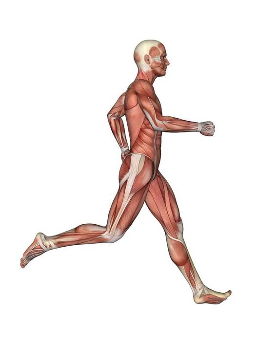
- It allows movement and locomotion, which differentiates us from plants and other sessile or immobile organisms, and which also characterizes us as human beings, capable of performing different conscious activities such as playing musical instruments, practicing sports, building things, dancing, draw, speak, sing, etc..
- Muscles function in processes essential for life such as the pumping of blood through the body, the movement and conduction of said blood, the movement of food, respiration, etc..
- They participate in the movement of the eyeballs, which allows us to have a wider range of vision.
- They help us move our jaws to chew food and swallow it (using the tongue), as well as to speak, cough, yawn, among others..
- They allow us to control excretory processes such as defecation or urination (urine), since the sphincters that control the openings to the outside of the tubes through which these wastes are expelled are made up of muscle tissues..
- They form a superficial layer that helps protect the internal organs of our body, especially against blows, accidents and others.
- Another important function has to do with the maintenance of posture, which depends on both the muscular and skeletal systems..
- Muscles also participate in the production of heat.
The muscular system is made up of muscle tissue. This tissue is made up of specialized cells called myocytes, which are also known as muscle fibers, in which resides the capacity for contraction or elongation that characterizes the muscles.
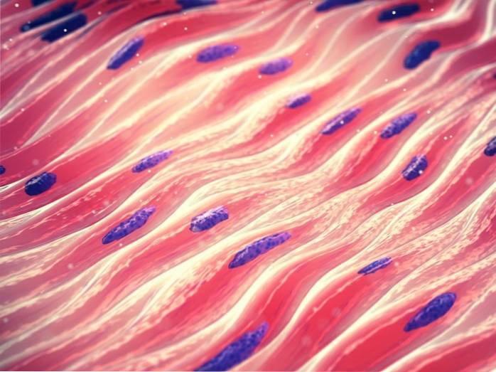
Although there are some differences between the different types of muscles, myocytes are generally elongated, fibrous-looking cells of great length (especially skeletal skeletal muscle cells)..
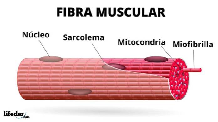
Inside we can find the organelles of any eukaryotic cell: plasma membrane, nucleus, mitochondria, endoplasmic reticulum and Golgi complex, lysosomes, peroxisomes and others..
However, some of these components are given special names, such is the case, for example, of:
The sarcolemma or the plasma membrane of muscle cells is a membrane similar to that of neuronal cells, which means that it can be excited and conduct action potentials, for which it is perfectly organized and arranged in myocytes..
In addition to the internal components common to all human cells, myocytes have within them a series of contractile elements called myofibrils, which are made up of contractile protein fibers called myofilaments..
There are two types of myofilaments that, together, are responsible for the shortening or lengthening of muscle tissues, these are the thick myofilaments and the thin myofilaments.
The thick myofilaments are made up of a protein called myosin II, while the thin filaments are mainly made up of protein actin F (which is a polymer of the protein G actin) and two other proteins that participate in the structure: troponin and the tropomyosin.
In addition to these, myofilaments are associated with other proteins that do not have contractile functions, but that play an important structural role, the two most important being titina and the nebulin.
Muscle cell contraction depends on the ability of thin myofilaments to slide over thick myofilaments, shortening the length of the cells they make up. In addition, it is a process that demands a constant supply of energy in the form of a molecule known as ATP..
Depending on how the myofilaments are structurally arranged in the myofibrils of muscle fibers or cells, two types of muscle cells are defined: striated and smooth.
These are the cells that make up the muscle tissue associated with the skeleton and heart muscles. Viewed under the microscope, these cells present a pattern of transverse bands or "striations" that are repeated throughout their entire structure, hence their name..
Myofibrils occupy practically the entire cell space of striated muscle fibers and are usually arranged longitudinally in parallel-ordered assemblages..
Unlike the previous ones, these muscle fibers do not present the pattern of transverse bands, which is why they are called “smooth fibers”.
The muscles of the intestines, blood vessels, sphincters, urinary bladder and other internal organs are made up of this type of cells..
They have a higher proportion of thin myofilaments relative to thick myofilaments, and these are longer than the thin myofilaments of striated myocytes.
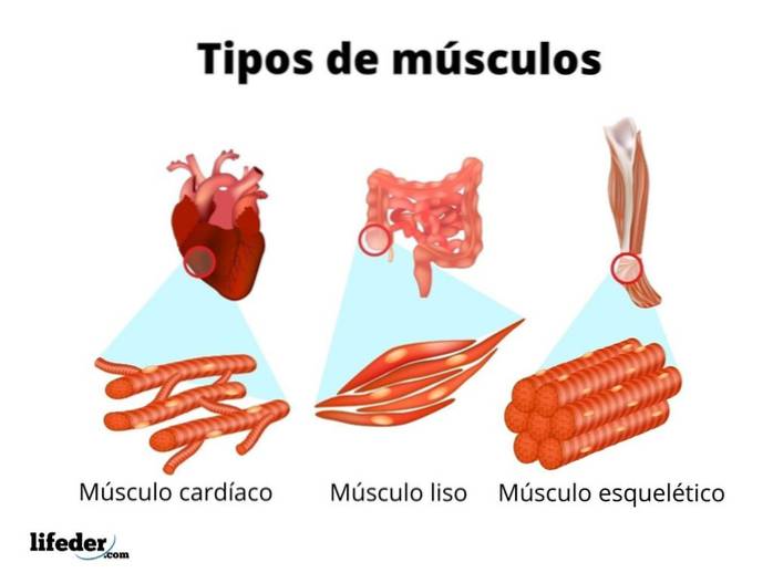
The main classification of the muscles of the human body is based on their histological characteristics and suggests the existence of three fundamental groups: skeletal muscle, cardiac muscle and smooth muscle..
Skeletal muscle is made up of striated muscle fibers, which are structurally and functionally separated from each other, but which contract in unison to allow movement..
These muscles associate with and join bones through connective tissue fibers known as tendons.
The contraction and relaxation of skeletal muscles is under the action of the central nervous system, specifically the somatic division of the peripheral nervous system, which controls our voluntary movements through motor neurons..
The heart is a muscular organ. It is made up of four hollow chambers with muscular walls that contract spontaneously and whose cells are regulated by neurons of the autonomic nervous system, making it an involuntary controlled organ..
Heart muscle cells are striated muscle cells or fibers that contain thick and thin myofilaments arranged in the same way as skeletal muscle cells..
However, they are shorter in length and interconnected, allowing the rapid propagation of electrical impulses that promote their contraction, no matter where they are generated..
This type of muscle is the one that forms the main hollow visceral organs of our body, such as the intestines (where they allow peristaltic movements that favor the digestive process), the blood vessels, the ureters and the uterus, among others. Smooth muscles are also those found in the iris of our eyes.
These muscles are under the control of the autonomic nervous system, which means that their contraction does not depend on our will, as is the case with skeletal muscle..
Depending on the organ to which they belong, smooth muscle cells are arranged in circular, longitudinal or both layers, which is closely related to their contractile functions..
These are the ones that allow us to have facial expressions, chew, speak, move the neck and head and breathe, they are skeletal muscles that are associated with the axial skeleton, which is the one that forms the skull, spinal column and rib cage..
The main muscles of the face are the following skeletal muscles:
- Occipitofrontal muscle: which is a large muscle located in the forehead.
- Temporal muscle- A thick fan-shaped muscle that allows the mouth to close and the jaw to move from side to side during chewing.
- Buccinator muscle: also called the "trumpeter muscle", it is a muscle found in the cheek and whose function is to prevent food from passing to the outer surface of the teeth when we chew.
- Masseter muscle: that goes from the cheek bone to the lower jaw and works in the approach and distance of the teeth when we eat, it is said to be the strongest of the muscles in our body.
- Chin muscle (mentalis): it is located on the chin and allows certain movements of the lower lip.
- Lower lip depressor muscle: associated with the lower lip, it allows to “lower” the lip when we make facial expressions of disgust.
- Orbicularis muscle of the mouth: a circular muscle around the lips that brings them closer to each other.
- Elevator upper lip muscle: located on both sides of the nose, it is the muscle that allows us to deepen the furrows on each side of the nose and upper lip when we make facial expressions of sadness.
- Orbicularis muscles of the eyes: that form the eyelids and orbitals, allowing us to open or close the eyelids when we blink.
- Laughter muscle: it is the muscle that allows us to stretch our lips horizontally, like when we fake a smile.

In addition to muscles the muscles of the face, other well-known muscles of the body, which are generally the focus of attention for those who go to the gym to train strength or to "define themselves" are the following:
- Biceps: they are the muscles found in the arms, exactly mechanically joining the arm with the forearm, where it fulfills the function of flexing the forearm.
- Triceps: it is also a muscle that is in the arm, but at the back of the arm, it represents a large percentage of the muscle mass of the arm.
- Pectorals: these are three muscles that are found in the chest region and that help us to make strength with the upper limbs.
- ABS: correspond to the set of muscles found in the abdominal area and that protect the organs and help keep the body straight and perform different movements.
- Abductors: they are responsible for the separation of the legs and is located in the hip region, between the thigh and the buttocks.
- Buttocks: they are the muscles that are in the back of the legs and make up the buttocks, they allow the movement of the thigh towards the rear.
- Quadriceps: it is a large muscle that is found in the front part of the thighs and that participates in the extension of the knee to make the leg straight.
The muscular system is prone to different diseases such as cancers, infections, wounds, trauma, etc..
Generally, diseases or disorders that have to do with skeletal muscles are classified into two categories: musculoskeletal disorders and neuromuscular disorders..
However, the heart and the viscera, as muscular organs, can also suffer from diseases of different kinds.
- Musculoskeletal disorders: injuries to muscles or tendons due to mechanical stress such as overextension, repetitive movements and poor posture. These include muscle tension, tendonitis, carpal tunnel syndrome, which are usually related to work or sports and affect few muscles.
- Neuromuscular disorders: these are systemic disorders that are related to problems of the nervous system to control the contraction of the muscles; they often have to do with genetic problems and affect a large number of muscles at the same time. Examples are muscular dystrophy and Parkinson's disease.
Taking care of our muscles is essential to keep them healthy and in good working order, so that we do not have to do with the consequences of their deterioration. Some helpful tips for taking care of the muscular system include:
- Always warm up the muscles before starting any physical activity and allow them to cool down after finishing it..
- Trying, as far as possible, to stretch most of the body's muscles on a daily basis, but especially before doing sports or any physical activity, this will prevent them from becoming fatigued too quickly or from prolonged use causing too much pain.
- Staying well hydrated, since water is very important for the functioning of our organs, for lubrication and heat dissipation.
- Have an adequate diet, where a mixture of fruits, vegetables, fats, carbohydrates and proteins of different types is consumed in a balanced way, always seeking to avoid excesses.
- Avoid the consumption of drugs of any kind, since they are harmful to health in general, either at the level of the nervous system (which will ultimately affect muscle functions) or at the systemic level in general..
- Exercising daily or at least 3-4 times a week, for a minimum of 30 minutes each time.
- Avoid stress and try to get enough sleep and rest.


Yet No Comments