Streptococcus agalactiae, Also known as Group B beta-hemolytic Streptococcus, it is a gram positive bacterium, the main cause of disease in the neonatal and perinatal periods. It is normally found as a common microbiota of the lower gastrointestinal tract, but from there it can colonize other sites, being able to be found in the female genital tract and in the pharynx.
The percentage of pregnant women who are carriers of the Streptococcus agalactiae it is 10% -40% and the transmission rate to newborns is 50%. Approximately 1-2% of colonized newborns will become ill from this bacteria.
In newborns, Streptococcus agalactiae it can cause septicemia, meningitis and respiratory infections, and in the mother it can cause puerperal infections and wound infection, among others.
This microorganism also behaves like an animal pathogen. It has been the main cause of bovine mastitis, interrupting the production of industrial milk, hence its name agalactiae, which means without milk..
Article index
S. agalactiae it is characterized by being facultative anaerobic, it grows well in media enriched with blood at 36 or 37 ºC for 24 hours of incubation. Their growth is favored if they are incubated in an atmosphere with 5-7% carbon dioxide.
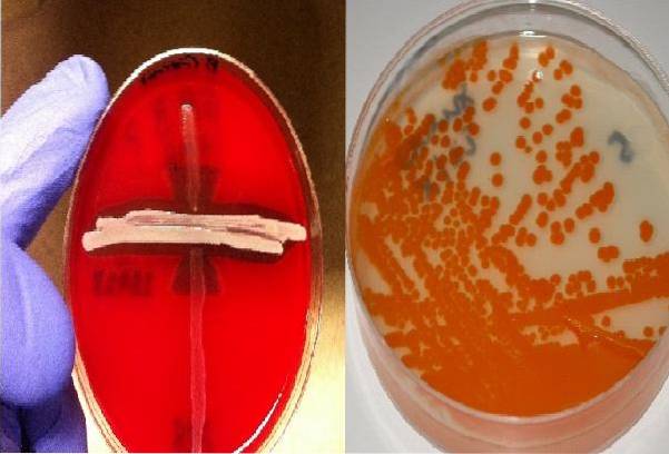
In blood agar they induce a halo of complete hemolysis around the colony (beta-hemolysis), thanks to the production of hemolysins, although the hemolysis produced is not as pronounced as that of other Streptococcus.
In New Granada agar it has the ability to produce a pathognomonic orange pigment of the species.
On the other hand, S. agalactiae it is catalase and oxidase negative.
Streptococcus agalactiae belongs to the Bacteria Domain, Phylum Firmicutes, Bacilli Class, Lactobacillales Order, Streptococaceae Family, Streptococcus Genus, Agalactiae Species.
Pbelongs to group B according to the Lancefield classification.
Streptococcus agalactiae are Gram positive cocci that are arranged as short chains and diplococci.
Slightly larger colonies can be observed on blood agar with less marked beta-hemolysis than that produced by Group A Streptococcus..
This microorganism has a polysaccharide capsule of nine antigenic types (Ia, Ib, II, - VIII). All have sialic acid.
Group B antigen is present in the cell wall.
The transmission of the bacteria from the mother to the child occurs mainly vertically. The child can be infected either in uterus, when the bacteria reach the amniotic fluid, or during the passage of the child through the birth canal.
The risk of transmission from mother to child is greater when there are predisposing factors. Among them are:
Although it has also been seen that it can be colonized by nosocomial exposure after birth.
The virulence mechanism exerted by this bacterium is aimed at weakening the defense systems of the patient to invade the tissues. Among the virulence factors is the capsule rich in sialic acid and beta hemolysin..
However, a variety of surface and extracellular matrix proteins that are capable of binding to fibronectin have also been identified..
In addition to this, sialic acid binds serum factor H, which accelerates the elimination of compound C3b from complement before it can opsonize the bacteria..
Of course, this renders the innate immunity line of defense through phagocytosis mediated by the alternate complement pathway ineffective..
Therefore, the only possible defense option is through complement activation by the classical pathway, but this has the disadvantage that it requires the presence of type-specific antibodies..
But for the newborn to possess this antibody, it must be provided by the mother through the placenta. Otherwise, the newborn is unprotected against this microorganism.
Besides this, S. agalactiae produces a peptidase that renders C5a useless, which results in very poor chemotaxis of polymorphonuclear leukocytes (PMN).
This explains why serious neonatal infections present with a low presence of PMN (neutropenia).
Generally the signs of infection in the newborn are evident at birth (12 to 20 hours after delivery up to the first 5 days) (early onset).
Nonspecific signs such as irritability, lack of appetite, respiratory problems, jaundice, hypotension, fever, or sometimes hypothermia begins to be observed.
These signs evolve and the subsequent diagnosis can be septicemia, meningitis, pneumonia, or septic shock, with a mortality rate in term-born infants of 2 to 8%, increasing considerably in premature infants..
In other cases, a late onset can be observed from day 7 of birth until 1 to 3 months later, presenting meningitis and focal infections in bones and joints, with a mortality rate of 10 to 15%.
Late-onset meningitis cases can leave permanent neurological sequelae in approximately 50% of cases..
From the mother's point of view, she may present with chorioamnionitis and bacteremia during peripartum.
You can also develop postpartum endometritis, post-caesarean section bacteremia, and asymptomatic bacteriuria during and after delivery..
Other affectations by this bacterium in adults can be meningitis, pneumonia, endocarditis, fasciitis, intra-abdominal abscesses and skin infections.
However, the disease in adults, even when serious, is not usually fatal, while in the newborn it is, with a mortality rate of up to 10% - 15%.
This microorganism can also affect older children, non-pregnant women, and even men..
These are generally weakened patients, where S. agalactiae can cause pneumonia with empyema and pleural effusion, septic arthritis, osteomyelitis, urinary tract infections, cystitis, pyelonephritis, and soft tissue infections ranging from cellulitis to necrotizing fasciitis.
Other rare complications can be conjunctivitis, keratitis and endophthalmitis..
The fetus can naturally be protected in the perinatal period. This is possible if the mother has IgG antibodies against the capsular specific antigen of the Streptococcus agalactiae of which it is colonized.
IgG-type antibodies are able to cross the placenta and this is how it protects it.
If, on the contrary, the IgG antibodies present in the mother are against another capsular antigen different to the type of S. agalactiae that is colonizing at the time, these will not protect the neonate.
Fortunately, there are only nine serotypes and the most frequent is type III..
However, obstetricians typically prevent neonatal disease by administering intravenous ampicillin to the mother prophylactically during labor..
This should be done whenever the mother has a positive vaginal sample culture for S. agalactiae in the third trimester of gestation (35 to 37 weeks).
However, this measure will only prevent early disease in the newborn in 70% of cases, having low protection over late-onset disease, since these are mostly caused by external factors post-birth.
In case the mother is allergic to penicillin, cefazolin, clindamycin or vancomycin can be used..
For diagnosis, the isolation of the microorganism from samples such as blood, CSF, sputum, vaginal discharge, urine, among others, is ideal..
It grows on blood agar and pomegranate agar. In both it has specific characteristics; beta-hemolytic colonies are observed in the first and orange-salmon colonies in the second.
Unfortunately, 5% of the isolates do not present hemolysis or pigment, so they would not be detected with these means.
Detection of capsular antigens from S. agalactiae in CSF, serum, urine and pure cultures it is possible by the latex agglutination method, using specific antisera.
Likewise, the CAMP factor detection test is very common to identify the species. It is an extracellular protein that acts synergistically with the ß-lysine of Staphylococcus aureus when sown perpendicular to S. agalactiae, creating a larger arrow-shaped area of hemolysis.
Other important diagnostic tests are the hippurate and arginine test. Both test positive.
It is efficiently treated with penicillin or ampicillin. Sometimes it is usually combined with an aminoglycoside because its administration together has a synergistic effect, in addition to increasing the spectrum of action in cases of infections associated with other bacteria.


Yet No Comments