
The capsule staining is a differential staining technique that has the property of highlighting the polysaccharide structure that surrounds certain bacteria and yeasts called capsule. It is used in clinical laboratories to help diagnose certain pathologies caused by capsulated microorganisms..
It is also used in teaching laboratories for the demonstration of this morphological structure to students of health sciences careers, such as: medicine, bioanalysis, nursing, or cytotechnology, among others..
There are several simple techniques to demonstrate the presence of the capsule in the microorganisms that possess it, these are: the negative stain, the Anthony stain and a variant that combines the previous two.
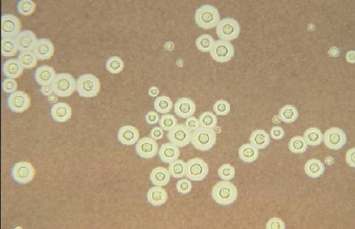
Negative staining is mainly used in CSF samples when the presence of yeast is suspected. Cryptococcus neoformans. This yeast is a common cause of meningitis.
This technique uses nigrosin or India ink and is based on creating a contrast between the background of the preparation and the impenetrable capsule of the microorganism. The background turns dark and the capsule is colorless. In this way, this structure is highlighted.
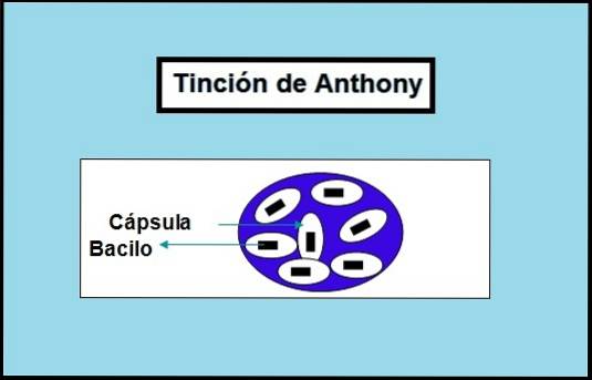
Regarding the Anthony technique, it can be said that it is mostly used in teaching laboratories to demonstrate the polysaccharide structure in bacteria such as Klebsiella pneumoniae, Streptococcus pneumoniae Y Neisseria meningitidis.
The use of this technique for diagnostic purposes is very rare, since there are other routine tests that allow the identification of these microorganisms..
Article index
The capsule is a strong structure of a polysaccharide nature. This protects microorganisms from phagocytosis, and therefore is a difficult structure to penetrate.
That is why capsule stains are based on contrast. The dyes stain the bottom of the preparation while the capsule remains colorless.
Therefore, with these techniques the capsule is easily recognizable. If the microorganism does not have a capsule, it will not be distinguishable with this type of coloration, because everything will be stained the same color..
All the techniques used to color the capsule have the same rationale despite using different colorants and procedures..
Anthony's stain uses crystal violet as a stain. This will stain the bacterial body and background purple.
On the other hand, 20% copper sulfate is used. This serves as a washing solution, that is, it removes excess violet crystal from the preparation, making the capsules clear but without losing the color of the bacterial body or the background..
- Iridescent milk.
- Microscope slide.
- 1% Violet Crystal.
- 20% copper sulfate.
- Optical microscope.
- Immersion oil.
This technique consists of:
It is important not to use heat neither to fix nor to dry, as this damages the capsule. Also do not wash with water.
Iridescent milk is an excellent culture medium as it provides the nutrients necessary for the microorganism to develop a prominent capsule.
On the other hand, the iridescent milk will form a thick and compact bottom that will be stained purple together with the bacterial body, but the capsule that surrounds the microorganism will remain colorless. Therefore, a clear halo is observed around the bacterial body..
It is a simple technique to perform. No fixing required.
Likewise, it should be noted that other culture media can be used, but the milk medium is preferred because it has the advantage that it provides more prominent capsules..
It is a slightly more laborious technique than negative staining, and its visualization requires waiting for the preparation to dry completely..
- Microscope slides.
- Culture medium with the microorganism.
- Chinese ink or nigrosine.
- Optical microscope.
- Physiological saline solution.
Place a drop of physiological saline solution on the coverslip and dissolve a small portion of the microbial culture. It is important that the preparation is not too thick. Then place a drop of Chinese ink or nigrosine and mix.
A coverslip sheet is then placed over the preparation without overflowing the liquid. It is observed under the microscope by first focusing on a 10X objective and then moving to 40X.
This technique can also be used on CSF samples directly. That is, instead of placing a drop of microbial culture, a drop of CSF is placed.
It is a simple method to perform and at the same time inexpensive. Does not require fixation or drying of the preparation.
It has the disadvantage that it must be observed under a microscope before the preparation dries, because if this happens the microorganisms will contract, which will make visualization difficult..
On the other hand, false positives can occur if the analyst is inexperienced, since leukocytes are often confused with yeasts..
The observation of yeast capsules with the Chinese ink or nigrosin technique should be considered as a presumptive diagnosis of Cryptococcus neoformans until it is demonstrated with the culture.
This is because there are other yeasts that could be the cause of meningitis and not just the Cryptococcus neoformans, such as those of the genus Candida and Rhodotorula, as well as other species of Cryptococcus.
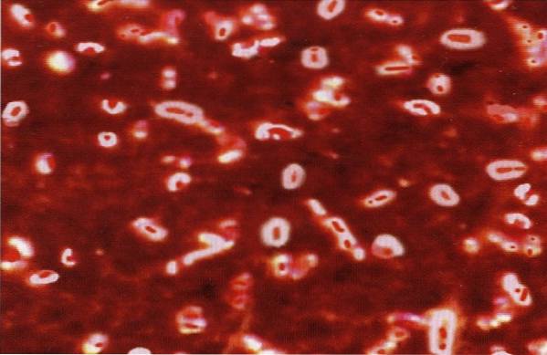
If there are capsulated microorganisms, a dark background will be observed, with transparent bodies floating in the liquid, highlighting the presence of the capsule..
This technique can also be done with nigrosine. It is a combination of the techniques previously explained. This technique uses crystal violet and Indian ink or nigrosine.
The bacterial body turns purple from the crystal violet because it is negatively charged, while nigrosin colors the bottom of the smear. If the bacterium has a capsule, it will appear as a transparent halo around the microorganism..
Crystal violet can be replaced by any of these colors: safranin, basic fuchsin or methylene blue.
- Violet glass.
- Nigrosine or Indian ink.
- Microscope slides.
- Microscope.
- Cultivate the microorganism in a culture medium.
- Place a drop of the culture on the end of a slide and next to it, place a drop of crystal India ink or nigrosin, mix and spread with the end of another slide..
- Air dry and do not heat set.
- Cover with a crystal violet solution for 1 minute, wash with distilled water but very delicately (gentle stream), allow to air dry.
- Observe under a microscope with an immersion objective. Search towards the ends of the spread.
A violet colored bacterial body and a dark background will be seen. The capsule, if present, will appear colorless around the bacteria.


Yet No Comments