The tropomyosin is one of the three proteins that are part of the thin filaments in the myofibrils of the muscle cells of the skeletal skeletal muscle of vertebrates and of the muscle cells of some invertebrates.
It is mainly associated with actin filaments in muscle myofibrils, but there are reports that indicate that, although to a lesser extent, it can also be associated with actin filaments in the cytoskeleton of non-muscle cells..
It was isolated and crystallized for the first time between 1946 and 1948, using protocols similar to those used years before to obtain actin and myosin, the two most abundant proteins in myofilaments..
In skeletal muscle cells, tropomyosin constitutes, together with troponin, a regulatory protein duo that acts as a calcium “sensor”, since its inhibitory association with actin fibers is reversed after binding with calcium ions that enter the cell in response to nerve stimuli that direct contraction.
Article index
In vertebrate cells, tropomyosin is invariably found as part of the thin filaments in muscle myofibrils, both in striated and smooth muscle, where it exerts regulatory functions..
Scientists have described tropomyosin as an asymmetric protein, quite stable against heat (thermostable), whose polymerization seems to depend on the ionic concentration of the medium where it is found..
It belongs to a large and complex family of fibrous and helical proteins that are widely distributed among eukaryotes. In vertebrates, tropomyosins are classified into two large groups:
- Those of high molecular weight (between 284-281 amino acids).
- Those of low molecular weight (between 245-251 amino acids).
All isoforms, when examined separately, have a number of amino acid residues that is a multiple of 40. There are hypotheses that each of these "clusters" of amino acids interacts with a G-actin monomer when both proteins are forming a complex. in the thin strands.
Mammals contain at least 20 different isoforms of tropomyosin, encoded by four genes that are expressed through alternative promoters and whose products (mRNA) are processed by alternative splicing (Splicing).
Some of these isoforms have differential expression. Many are tissue and stage-specific, as some are found in specific muscle tissues and it may be the case that they are expressed only at a specific time in development..
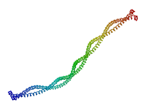
Tropomyosin is a dimeric protein, composed of two coiled alpha polypeptide helices, of more or less 284 amino acid residues each, with a molecular weight close to 70 kDa and a length of more than 400 nm.
Since there can be multiple isoforms, their structure can be composed of two identical or two different molecules, thus forming a homodimeric or heterodimeric protein, respectively. They differ in the "strength" with which they bind to actin filaments..
Tropomyosin molecules, also filamentous in shape, are located in the "groove" regions that exist between the G-actin polymer chains that make up the F-actin strands of fine filaments. Some authors describe their association as a "form complementarity" between both proteins..
The sequence of this protein is conceived as a "string" of repeating heptapeptides (7 amino acids), whose individual characteristics and properties promote the stable packaging of the two helices that make up its structure, and between which the binding sites are formed. for actin.
The union between tropomyosin fibers and actin fibers occurs mainly through electrostatic interactions.
The N-terminal end of tropomyosins is highly conserved among the different muscle isoforms. So much so, that eight of the first nine residues are identical from man to Drosophila (the fruit fly), and 18 of the first 20 N-terminal residues are conserved in all vertebrates.
Tropomyosin and troponin, as discussed above, constitute the regulatory duo of muscle contraction of skeletal and cardiac fibers in vertebrates and some invertebrates..
Troponin is a protein complex made up of three subunits, one that responds to and binds to calcium, another that binds to tropomyosin, and another that binds to F-actin filaments.
Each tropomyosin molecule is associated with a troponin complex that regulates the movements of the first.
When the muscle is relaxed, tropomyosin is in a special topology that blocks myosin-binding sites on actin, preventing contraction.
When muscle fibers receive the appropriate stimulus, intracellular calcium concentration increases, causing a conformational change in troponin associated with tropomyosin.
The conformational change in troponin also induces a conformational change in tropomyosin, which results in the "release" of the act-myosin binding sites and allows contraction of the myofibrils to occur..
In non-muscle cells where it is found, tropomyosin apparently fulfills structural functions or in the regulation of cell morphology and mobility..
Tropomyosin has been identified as one of the most abundant allergenic muscle proteins in cases of allergic reactions caused by foods of animal origin..
It is present in muscle and non-muscle cells, both vertebrates and invertebrates. Various studies reveal that allergic reactions caused by crustaceans such as shrimp, crabs and lobsters are the product of the "detection" of their epitopes by means of immunoglobulins in the serum of hypersensitive allergic patients..
This protein is thought to behave as a cross-reactive allergen, since patients allergic to shrimp, for example, are also allergic to other crustaceans and mollusks that have a protein with similar characteristics.

Yet No Comments