
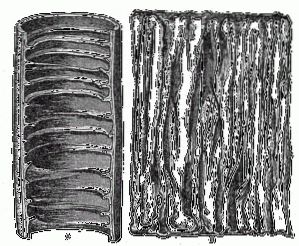
The conniving valves, Kerckring folds, or simply circular folds, are permanent folds found in the mucosa and submucosa of the small intestine. These are transverse folds that form macroscopic helical or semicircular elevations, which can encompass the entire internal perimeter of the digestive tract..
Along with intestinal villi and microvilli, conniving valves are one of the most important modifications found on the luminal surface of the small intestine..
These are particularly abundant in the portions of the intestine known as the duodenum and jejunum, that is, in the first two portions of this section of the digestive tract, and they decrease to the most distal portions of the ileum..
Their main function is to increase the surface area for the absorption of nutrients during the digestion of food, however, they also seem to participate in the flow of chyme (pre-digested food mass in the mouth and stomach)..
They were discovered by the German anatomophysiologist Thomas Theodor Kerckring in 1958, for whom they were named.
However, those arranged transversely to the longitudinal axis of the intestine are also called "connivent valves", since they narrow or reduce the diameter of the intestinal lumen, although they do not behave like true valves..
Article index
The small intestine, which is a tube approximately 7 meters long, is divided into three segments: the duodenum, jejunum, and ileum. The duodenum is the intestinal portion that connects to the stomach and does so through the pyloric region.
The duodenum is continued with the jejunum and the latter with the ileum. In turn, the ileum is the connection of the small intestine with the large intestine, through the ileocecal valve.
If a cross section of the wall of the small intestine is observed, 5 well-defined layers can be seen. These are known as the mucous layer, the submucosa, the circular muscle, the longitudinal muscle, and the serosa..
Of all these, the mucosa is the one that presents specializations that allow it to considerably increase its surface area..
Of these specializations, the most prominent are the connivent valves, which are very abundant in the upper portions of the intestine and diminish as we approach the final portions of the ileum..
Kerckring circular folds can be 3 to 10 mm high and up to 5 cm long, and are distributed at regular intervals every 3 cm. In the intestine of a normal adult, these can be found in a number that ranges between 400 and 900.
Pathological studies have shown that the average length of the mucosal valves in the unfolded state is around 14 meters, while in the valvular state of the mucosa this length is more or less half (the 7 meters of the intestine).
Not all the folds seen in the intestine cover the entire perimeter of the tube. Some Kerckring valves extend around the entire circumference, while others only span two-thirds of the circumference or less (crescent valves), and others may spiral several times around the circumference..
They have been called “valves” due to their ability to reduce luminal space, but the qualifier “connivent” responds to their permissive nature in both directions, since they are not occlusive valves..
Each fold is richly vascularized and receives a large network of lymphatic vessels. Both types of vessels run within each other through the submucosal layer, which is immediately below the mucosa..
The surface of each fold is covered with intestinal villi and these, in turn, have microvilli, which gives it a characteristic velvety appearance.
Conniving valve distribution and conformation abnormalities are associated with some cases of partial or complete intraluminal obstruction of the small intestine. Alterations in the orientation of these structures have been related to some diseases of the small intestine.
The most important function of Kerckring valves is, without a doubt, to provide a huge surface area for the absorption of nutrients, in addition to supplying the functional structures for this purpose..
In other words, all the functions of these permanent structures present in the intestinal lumen are directly related to the functions of the other surface modifications associated with them, such as villi and microvilli..
Together, the presence of Kerckring folds, villi, and microvilli achieves up to a 600-fold increase in the total surface area of a smooth tube.
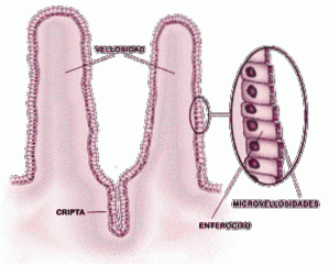
Since the villi that cover these valves contain a great variety of cells with secretory and absorption functions, we can refer to the digestive and absorption functions of the conniving valves..
Enterocytes (cells in the intestine) perform different absorption functions throughout the small intestine..
The duodenum absorbs mainly iron, calcium, lipids, sugars, water, proteins, vitamins, magnesium and sodium. The cells present on the luminal surface of the jejunum are responsible for the absorption of sugars and proteins, mainly.
Finally, bile salts, vitamin B12 and chlorine ions are reabsorbed in the ileum..
Few diseases have been directly related to conniving valves, beyond those concerned with malformations or congenital defects in their development..
However, since they are permanently exposed to contact with potential pathogens, these mucosal structures can suffer infection, injury, inflammation and growth..
As mentioned, some conditions related to intestinal obstructions may be due to edema or thickening of the folds of the mucosa..
Examples of pathologies of this type are lymphomas and regional enteritis, characterized by malabsorption processes in the small intestine, caused by thickening of the Kerckring folds..
Whipple's disease, in 80% of cases, is due to the presence of prominent folds in the duodenum and jejunum region, in addition to the proliferation of macrophage-like cells within the lamina propria of the small intestine.

Yet No Comments