Chilomastix mesnili it is a protozoan that belongs to the group of flagellates. Parasites the digestive tract of humans, specifically the colon, without causing any type of pathology.
He was first observed by the French physician Casimir Devine, who first named him Cercomonas intestinalis. Subsequently, the genre was created Chilomastix to include organisms with these characteristics.
The Chilomastix mesnili it is a very well known protozoan, which has been sufficiently studied, especially its characteristics and behavior within the human intestine. Because of this, it has been established that he poses no threat to his host..
Article index
The taxonomic classification of Chilomastix mesnili is the next:
Chilomastix mesnili it is an organism that belongs to the group of flagellate protozoa. It has 4 flagella, three of which contribute greatly to its locomotion.
This protozoan is commonly found in the large intestine of some primates such as humans. Specifically, it is housed in the cecum, the first portion of the large intestine where the appendix is also found..
In this type of organism, only the asexual type of reproduction is observed, which does not require the fusion of sex cells.
The asexual reproduction method of the Chilomastix mesnili is the binary fission. In this process, the first thing that happens is DNA duplication. Subsequently, the cytoplasm of the cell divides following the longitudinal plane, originating two cells, each of which is exactly the same as the cell that gave rise to them..
Chilomastix mesnili it is a heterotrophic organism, which means that it is not capable of synthesizing its own nutrients.
It feeds through phagocytosis, a fairly common process in protozoa. Through this process, food particles from the digestive tract enter the cell through the cytostome to be processed and assimilated.
In general, the Chilomastix mesnili It is a protozoan that does not represent any risk to the health of its host, since it does not cause imbalances at the intestinal level.
On very rare occasions it can cause some discomfort, this being related both to the number of parasites in the intestine, and to the immune status of the host..
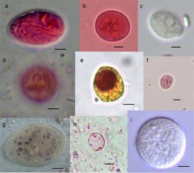
The Chilomastic mesnili, During its life cycle it can be found in two forms: cyst and trophozoite. Each one has a totally different morphology.
It represents the infectious form of this protozoan. Seen under the microscope, it can be seen that the cysts are uninucleated, that is, they have a single nucleus. This nucleus is large, compared to the size of the cyst, occupying a large part of it. They are surrounded by a thick and resistant wall.
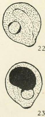
It has an oval shape, similar to a pear or a lemon and an anterior hyaline protrusion is observed on its surface. They measure on average between 6-10 microns long by 4-6 microns wide. They are also colorless. They do not present cytostoma or flagella.
It is the vegetative form of the protozoan, that is, the one that reproduces and feeds. It is pear-shaped. It measures approximately 11-16 microns. The cytoplasm is prominent, surrounded by microfibrils. It also lacks mitochondria and the Golgi apparatus. It has a spherical nucleus that measures between 3-4 microns on average; this is not visible when fresh preparations are made.
Likewise, in the microscope it is possible to observe the presence of several flagella (4), one of them being associated with the cytostome, which is a kind of opening through which the food particles enter the protozoan.
The trophozoite has a characteristic rotary motion.
The life cycle of Chilomastix mesnili it is monoxenic. In this type of cycle, the parasite needs only one host for its full development. In the case of this protozoan, the host is the human being.
It is important to note that this protozoan is a commensal of the large intestine of humans and other primates. This means that it lives there, at the expense of the bacteria that are part of the bacterial flora, but without causing any kind of damage to the host..
It is at the level of the cecum (large intestine) where trophozoites develop, reach their adult stage and reproduce, generating cysts. It is important to remember that these are the infecting form of the parasite..
The cysts are expelled to the external environment as part of the feces, mainly those that are well formed. In semi-liquid stools, both cysts and trophozoites have been observed. In those of the liquid type, the most frequently observed parasitic form of this protozoan are the trophozoites..
When they are ingested by some other host, they again lodge in the large intestine, where they proceed with their development until they become trophozoites and again generate other cysts, thus continuing the biological cycle..
The Chilomastix mesnili it is a protozoan considered harmless for humans. However, when its levels in the large intestine rise, it is possible that it can cause some intestinal-type discomfort.
The most common form of transmission is from person to person through the fecal oral mechanism. This involves the ingestion of food or water contaminated by fecal particles with parasitic cysts..
The Chilomastix mesnili it is a protozoan that regularly inhabits the large intestine of approximately 15% of the world's population. In general, it is not pathogenic, that is, it does not cause any type of damage or discomfort.
However, on certain occasions, when the number of parasites increases abnormally, a clinical picture compatible with a diarrheal-type intestinal infection is likely to occur. Among the symptoms that have been observed most frequently are:
As in any intestinal parasitic infection, the first diagnostic method is a stool examination, in which the infecting forms (cysts) of the parasite can be visualized through the microscope..
Importantly, performing a single negative test does not exclude infection. This is why it is necessary to carry out serial examinations to increase sensitivity and thus reach an accurate diagnosis..
Likewise, there are other techniques that seem to have a greater sensitivity in this type of diagnosis. These include:
It is a sedimentation-type procedure that is based on the use of low-density liquids. Through this process, it is possible to recover the parasitic cysts that are deposited at the bottom of the container because their density is greater. In this method, the combination of formaldehyde / ether or methyl acetate can be used as reagents..
This method uses zinc sulfate as a reagent. As this substance has a higher density than the water that is mixed with the feces, it allows the parasitic forms (cysts, eggs or larvae) to float and in this way they can be identified with the help of the microscope.
Taking into account that Chilomastix mesnili it is a parasite that in the vast majority of cases does not cause any type of pathology in man, there is no specific treatment to treat it.
However, in those cases in which it triggers any symptoms, doctors decide on drugs that have a clear broad-spectrum antiparasitic effect, such as metronidazole..
Prevention methods are the same as for other diseases caused by intestinal parasites. These consist of avoiding contamination caused by feces containing parasitic forms. Among the most relevant and common measures is washing your hands after going to the bathroom and before preparing any food.


Yet No Comments