
The kinetochore it is a protein complex associated with the centromeres of chromosomes in higher eukaryotes. It represents the main point of attachment for the microtubules of the spindle during cell division, either by mitosis or by meiosis.
Eukaryotic chromosomes have a special region known as the centromere, which is actually a very compact segment of DNA (in the form of chromatin), whose main function is to ensure the proper distribution of duplicated chromosomes during cell division..
During mitosis, for example, the genetic material (DNA) of a cell is duplicated during interface, resulting in the formation of two copies of each chromosome, which condense during the metaphase and are visualized as two sister chromatids, joined together through the centromere.
The separation of these chromatids towards each pole of the cell when division begins occurs thanks to the adhesion of the microtubules of the mitotic spindle to the centromeric region, specifically to the protein complex associated with it, known as the kinetochore..
Each chromosome is associated with two kinetochores, to which are attached microtubules of the mitotic spindle known as kinetochoric microtubules. This union occurs thanks to the connection between said microtubules and protein fibers that emerge from the kinetochore.
Article index

Kinetochores are formed in the centromeric region of chromosomes after their duplication, which takes place in the stages prior to cell division.
This formation depends on the association of the kinetochore with special portions of DNA in the region of the centromere, which serve as a scaffold for the formation of the former..
In these regions, the nucleosomes that compact the centromeric DNA in the form of chromatin are formed with a special variant of histone H3.
There are some differences in the structure of the kinetochore between plant and animal cells, and it is the kinetochore of the cells of mammalian animals that have been most extensively studied..
In general terms, it is said that the kinetochore protein complex has a "laminar" structure, with an internal region and an external one, the first specialized in the union of proteins to chromosomal DNA and the second in the union of the spindle fibers..
Some specialists in the field highlight the presence of a third "sheet" or "layer", which represents the interface between the internal and external regions..
The internal region of the kinetochore is formed by a constitutive network of proteins associated with the centromere, known as CCAN (from the English Constitutive Centromere-Associated Network), many of which are directly associated with the histone proteins of the centromeric nucleosomes.
The outer region of the kinetochore, on the other hand, is made up of two main protein complexes known as the Ndc80 complex and the Mis12 complex, each made up of several protein subunits..
Of both, Ndc80 is essential for the formation of microtubule binding sites and Mis12 is the “bond” between the components of the inner and outer regions of the kinetochore..
The formation of kinetochores in eukaryotic chromosomes depends on more than 50 proteins (some authors propose that more than 100), and it is during this process that the internal and external regions of these structures become evident and are formed..
The main point of reference for the formation of the kinetochore on the chromosomal centromeres is the variant of histone H3 known as Cse4 / Cnp1 / CENP-A, since it is necessary for the localization of almost all the proteins of the kinetochore..
We can say that the assembly process of this complex requires the specific recognition of multiple participants, each one with specific functions and probably in a sequence or hierarchical order:
- Some proteins function in the recognition of the parts involved, that is, the centromeric nucleosomes and the microtubules of the spindle..
- Some proteins work in stabilizing the protein complexes around the centromere.
- Others participate in stabilizing the junctions between microtubules and the kinetochore..
- There are proteins that prevent the separation of the chromatids until the kinetochores are perfectly attached to the mitotic spindle from each cell pole..
- There are also proteins that couple the movement of chromosomes with the depolymerization of spindle microtubules..
- These complexes also include motor proteins such as the dynein / dynactin pair that, among other things, function in the recruitment of regulatory proteins to the kinetochore and in the movement of chromatids..
- Finally, there are proteins that regulate the function of the other proteins in the complex, inhibiting or promoting their activity..
The kinetochore is a very important part complex associated with the centromere because, as we have commented, the correct segregation or separation of sister chromatids during cell division depends on it..
The segregation of these chromatids is essential for the maintenance of cell life, since each daughter cell must receive the same amount of genetic material during the division of the cell that gives rise to it, in order to perpetuate the cell line and / or the organism in question.
In addition to this function, many authors suggest that the kinetochore functions as an organizing center for the microtubules that are directed towards the chromosomes..
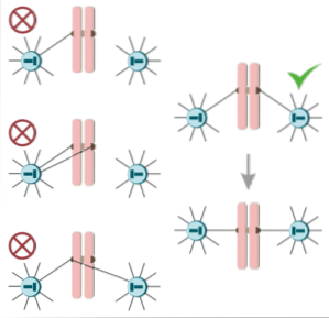
Cell division, whether by mitosis or meiosis, is a delicate process that requires great care and rigor, which is evidenced by the existence of what are known as "control points".
One of these checkpoints involves the cell "making sure" that the mitotic spindle fibers are correctly attached to the chromosomes through the kinetochores. Fibers from opposite poles of the dividing cell should be attached to each of the sister chromatids, in order to separate them properly.
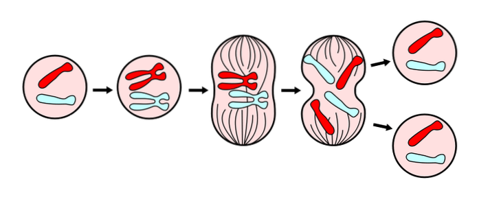
When the chromosomes have been correctly duplicated, the kinetochores and the fibers of the mitotic spindle function in the ordering of the chromosomes and their copies in the central region of the cell (also known as the metaphase plate)..
During the anaphase, When the spindle fibers "pull" each copy of the chromosomes to opposite poles of the cell, then some of the kinetochoric proteins that hold the sister chromatids together are disassembled, allowing their separation..
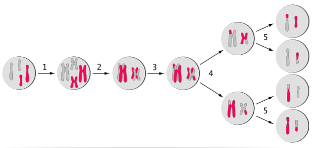
Meiosis is a process of cell division very similar and at the same time very different from mitosis, since the cell divides "twice".
During the first meiotic division, the kinetochores attach to the spindle fibers that come from each pole, separating the homologous chromosomes and not the sister chromatids..
Later, during the second division, the kinetochores are again connected to the fibers of the spindle that arise from each pole, separating the sister chromatids for their distribution among the daughter cells..
The success of the production of “healthy” sex cells depends, to a great extent, on the correct function of the kinetochores on each chromosome, since the wrong segregation of any chromosome can cause important pathological conditions in humans, such as trisomy 21 or Down syndrome, for example.


Yet No Comments