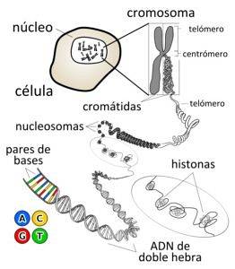
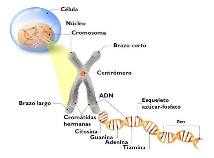
The chromosomes They are structures made up of a continuous DNA molecule and associated proteins. They are neatly found within the nucleus of eukaryotic cells and contain most of their genetic material. These structures are most clearly seen during cell division.
Eukaryotic chromosomes were first identified and studied in the late 18th century. Today the word "chromosome" is a widely known term, even for people who have studied only the most elementary aspects of biology or genetics..
On chromosomes are genes, many of which code for proteins, enzymes, and the information necessary for the life of each cell. However, many chromosomes fulfill purely structural functions, which means that they allow a specific arrangement of genes within the nuclear interior..
Generally, all the cells of an individual have the same number of chromosomes. In humans, for example, each of the trillion cells that have been estimated to make up the adult body has 46 chromosomes, which are organized into 23 different pairs.
Each of the 46 chromosomes in humans and other living organisms has unique characteristics; only those known as "homologous pairs" share characteristics with each other, but not with different pairs; that is, all chromosomes 1 are similar to each other, but these are different from 2 and 3, and so on.
If all the chromosomes of a human cell were arranged in a linear way, they would form a chain of more or less 2 meters in length, so one of the main functions of chromosomes is to compact the genetic material so that it "fits" in the nucleus, while allowing access of the transcriptional and replication machinery.
Despite the enormous differences that exist between bacterial genomes and those of eukaryotic organisms, the genetic material of prokaryotes (as well as that of some internal organelles of eukaryotes) is also called a chromosome and consists of a circular molecule.
Article index
At the time that Mendel determined the principles of heredity, he had no idea of the existence of chromosomes. However, he concluded that heritable elements were transmitted in duplicate through special particles, a notion well ahead of its time..
Two 18th century scientists, the botanist K. Nageli and the zoologist E. Beneden, engaged in the observation and study of plant and animal cells during cell division events; These were the first to describe structures shaped like "little rods" inside the central compartment known as the nucleus..
Both scientists detailed that, during the cell division of a "typical" cell, a new nucleus was formed, within which a new set of "small rods" appeared, similar to the one found in the cell initially..
This division process was later described with more precision by the German scientist W. Flemming in 1879, who, using dyes during observation, managed to stain the "little rods" to better visualize them..
T. H. Morgan demonstrated that phenotypes are inherited in the manner suggested by Mendel and that the units of inheritance reside on chromosomes. Morgan provided the physical evidence that consolidated the "Mendelian Revolution".
Flemming documented the behavior of the "rods" during interphase and cytokinesis (cell division). In 1882 he published an investigation where he first coined the term "chromatin" for the substance that stained inside the nucleus when the cell was not in division..
He had also observed that during cell division the number of "rods" (chromosomes) in the nucleus doubled. One of each pair of duplicated chromosomes was housed within each nucleus of the resulting cells, so the chromosomal complement of these during mitosis was identical.
W. Waldeyer, following the works of Flemming, established the term "chromosome" (from the Greek "body that stains") to describe the same substance that was arranged in an orderly manner at the time of cell division..
Over time, different researchers delved into the study of genetic material, with which the meaning of the terms "chromosome" and "chromatin" changed a bit. Today a chromosome is a discrete unit of genetic material and chromatin is the mixture of DNA and proteins that composes it..
E.B. Wilson, in the second edition of the book The cell (The Cell) established the first classification of chromosomes, which is based on the location of the centromere, a characteristic that influences the attachment of chromosomes to the mitotic spindle during cell division.
There are at least three different ways to classify chromosomes, since there are different chromosomes between species and in individuals of the same species there are chromosomes with different structures and functions. The most common classifications are:
The genetic material inside bacteria is seen as a dense and ordered circular mass, while in eukaryotic organisms it is seen as a dense mass that appears "disorganized" inside the nucleus. Depending on the cell, chromosomes can be classified into two large groups:
- The prokaryotic chromosomes: each prokaryotic organism has a single chromosome composed of a covalently closed (circular) DNA molecule, without histone proteins and located in a region of the cell known as the nucleoid.
- The eukaryotic chromosomes: in a eukaryote there may be two or more chromosomes for each cell, these are located inside the nucleus and are more complex structures than the bacterial chromosome. The DNA that makes them up is highly packed thanks to its association with proteins called "histones".
The centromere is a portion of the chromosomes that contains a fairly complex combination of proteins and DNA and that has a primary function during cell division, as it is responsible for “making sure” that the chromosome segregation process occurs..
According to the structural location of this "complex" (the centromere), some scientists have classified chromosomes into 4 categories, namely:
- Metacentric chromosomes: These are those whose centromere is in the center, that is, where the centromere separates the chromosomal structure into two portions of equal length.
- Submetacentric chromosomes: chromosomes where the centromere is deviated from the "center", contributing to the appearance of an "asymmetry" in length between the two portions that it separates.
- Acrocentric chromosomes: in acrocentric chromosomes, the centromere "deviation" is considerably marked, resulting in two chromosomal sections of very different sizes, one very long and one truly short..
- Telocentric chromosomes: those chromosomes whose centromere is located at the ends of the structure (telomeres).
Organisms that have sexual reproduction and that have separate sexes have two types of chromosomes that are classified, according to their function, into sex chromosomes and autosomal chromosomes.
Chromosomes autosomal (or autosomes) participate in the control of the inheritance of all the characteristics of a living being, except for the determination of sex. Humans, for example, have 22 pairs of autosomal chromosomes.
Chromosomes sexual, As their name indicates, they fulfill an elementary function in determining the sex of individuals, as they carry the necessary information for the development of many of the sexual characteristics of females and males that allow the existence of sexual reproduction..
The main function of chromosomes, in addition to housing the genetic material of a cell, compacting it so that it can be stored, transported and “read” within the nucleus, is to ensure the distribution of genetic material among the cells resulting from division..
Why? Because when chromosomes separate during cell division, the replication machinery faithfully "copies" the information contained in each DNA strand so that the new cells have the same information as the cell that gave rise to them..
Furthermore, the association of DNA with the proteins that are part of chromatin allows at the same time the definition of a specific “territory” for each chromosome, which is of great importance from the point of view of gene expression and identity. mobile.
Chromosomes are considerably far from being static or "inert" molecules, in reality it is the opposite, histone proteins, which are those that collaborate with the compaction of each DNA molecule in a chromosome, also participate in the dynamism that has to do with the transcription or silencing of specific parts of the genome.
Thus, the chromosomal structure not only works on the organization of DNA within the nucleus, but also determines which genes are “read” and which are not, directly influencing the characteristics of the individuals who carry it..
The structure of a chromosome can be analyzed from a “microscopic” (molecular) point of view and from a “macroscopic” (cytological) point of view..
A typical eukaryotic chromosome is made up of a linear double-stranded DNA molecule that can be hundreds of millions of base pairs in length. This DNA is highly organized at different levels, which allows it to be compacted.
The DNA of each chromosome is initially compacted by its "winding" around an octamer of histone proteins (H2A, H2B, H3 and H4), forming what is known as a nucleosome, which is 11 nanometers in diameter.
The association between histone proteins and DNA is possible thanks to an electrostatic interaction, since DNA is negatively charged and histones are basic proteins, rich in positively charged amino acid residues.
One nucleosome connects to another through a junction region formed by part of the DNA strand and by a histone protein, H1. The structure resulting from this compaction looks similar to a string of beads and decreases the length of the DNA strand by about 7 times..
DNA is further compacted when chromatin (DNA + histones) in the form of nucleosomes coils on itself, forming a fiber approximately 30 nm in diameter, which compacts the DNA strand another 7 times,
The 30 nm fiber is associated, in turn, with the filamentous proteins of the nuclear matrix (the laminae), which line the inner surface of the inner nuclear membrane. This association allows the progressive compaction of the fiber, since "loop domains" are formed that are anchored to the matrix, organizing the chromosomes in defined regions inside the nucleus..
It is important to note that the level of compaction of chromosomes is not equal throughout their entire structure. There are places that are hyper compacted, which are known as heterochromatin and which are generally "silent" genetically speaking..
The looser or more relaxed sites of the structure, those to which the replication or transcription machinery can access with relative ease, are known as euchromatic sites, being regions of the genome transcriptionally active.
When the cell is not dividing, the chromatin is seen as "loose" and even "disordered". However, as the cell cycle progresses, this material condenses or compacts and allows the visualization of the chromosomal structures that are described by cytologists..
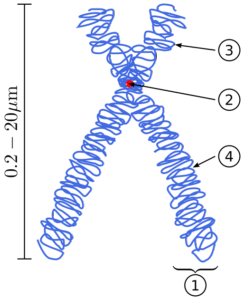
During the metaphase of cell division, each chromosome is seen as composed of a pair of cylindrical "chromatids" that are linked together thanks to a structure known as the centromere..
The centromere is a very important part of chromosomes, as it is the site to which the mitotic spindle attaches during division. This union allows the chromatids that are attached through the centromere to be separated, a process after which they are known as "daughter chromosomes.".
The centromere consists of a complex of proteins and DNA that is shaped like a "knot" and its location along the structure of a chromatid directly influences the morphology of each chromosome during nuclear division..
In a specialized region of the centromere is what scientists know as the kinetochore, which is the particular site where the mitotic spindle joins to separate sister chromatids during cell division..
The position of the centromere also determines the existence of two arms: a short or small one (p) and a larger one (q). In view of the fact that the position of the centromeres is practically unchanged, cytologists make use of the nomenclature "p" and "q" during the description of each chromosome..
These are specialized DNA sequences that "protect" the ends of each chromosome. Its protective function is to prevent different chromosomes from joining each other through their ends.
These regions of the chromosomes have received great attention, since scientists consider that telomeric sequences (where DNA forms structures somewhat more complex than a double helix) influence the activity of surrounding genes and, in addition, in the determination of the longevity of a cell.


Yet No Comments