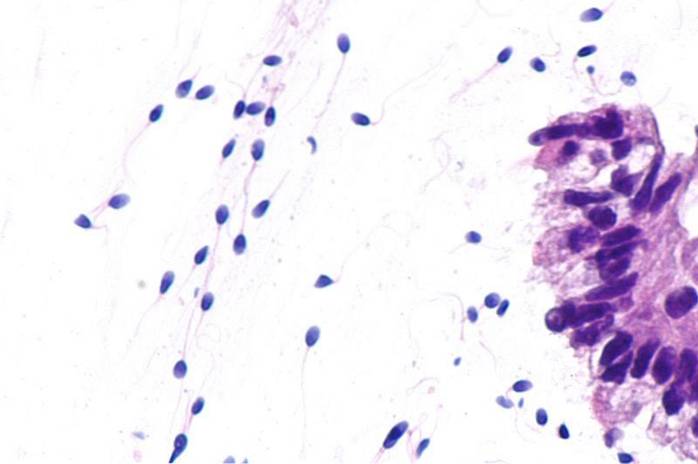
A primary spermatocyte It is an oval cell that is part of spermatogenesis, a process that results in the production of sperm. Primary spermatocytes are considered the largest cells of the seminiferous epithelium; possess 46 chromosomes and duplicate their DNA in the interphase process.
To reach the formation of a primary spermatocyte, the formation of a cell type called spermatogonia must occur in the testes. Upon entering prophase I, it becomes a primary spermatocyte that continues the process of reductive mitosis (first meiotic division).
Spermatocytes must reduce their chromosomal load in order to become the final gamete with 23 chromosomes. Primary spermatocytes enter a prolonged prophase of about 22 days and give rise to secondary spermatocytes; These originate the spermatids, which mature and become spermatozoa ready to fertilize.
The global gametogenesis process lasts about 74 days and involves a diploid spermatogonia that divides and finally forms four haploidly charged spermatozoa. A man can form an average of 300 million sperm a day.
Article index
Primary spermatocytes are the largest germ cells that can be found in the seminiferous tubules, in the intermediate layers of the germ epithelium. They come from the cell division of spermatogonia.
Morphologically they do not have any similarity to the mature sperm, made up of a head and a typical flagellum that gives it mobility. In contrast, they are oval cells that have the ability to grow continuously due to the accelerated manufacture of proteins, organelles, and other cellular products..
With regard to cellular behavior, the cytoplasm in these cells contains a greater amount of endoplasmic reticulum than spermatogonia. Similarly, the Golgi complex is more developed.
Spermatocytes can be differentiated from spermatogonia since they are the only cell type in which meiosis processes occur..
The cytokinesis process is particular, since the resulting cells form a syncytium and remain united by a cytoplasmic portion of 1 µm in diameter that allows communication between them and the exchange of certain molecules, such as proteins..
The spermatogenesis process occurs in the seminiferous tubules and is made up of two cell types: germ cells or spermatogonia and Sertoli cells..
The formation of primary spermatocytes was described by Erwing et al. In 1980, and in humans by Kerr and de Krestser in 1981.
Spermatogonia are the cells that give rise to the primary spermatocyte. These are fairly thick cells, with a round shape and homogeneous cytoplasm. They can be classified according to the morphology of their nucleus into: elongated type A, light type A, dark type A and type B.
Type A spermatogonia are stem cells and have reserve functions. A group of type A spermatogias differentiate and produce those of type B, which after multiple divisions give rise to primary spermatocytes.
As spermatogenesis progresses, the primary spermatocyte increases in size and notable changes can be seen in the morphology of the nucleus. Spermatocytes are able to migrate when the junctions between Sertoli cells disappear.
Sertoli cells are involved in the regulation of the entire spermatogenesis process. They are lining the seminiferous tubules and their function is to nourish the germ cells, give them support, serve as a barrier between the interstitium and the germ cells and mediate the cellular metabolic exchange.
Similarly, hormonal regulation occurs mainly in Sertroli cells, which have receptors for testosterone and FSH (follicle stimulating hormone)..
When activation by FSH occurs, a large number of key proteins are triggered so that this process can occur, vitamin A and ABP, among others.
Primary spermatocytes, which have a diameter of 16 mm, reach the middle of the germ tissue and undergo meiotic division to divide their chromosomal load. Now each daughter cell is called a secondary spermatocyte..
Secondary spermatocytes are also rounded but smaller cells. These cells undergo rapid meiotic division resulting in spermatids.
In other words, once meiosis I (reductional meiosis) is finished, meiosis II (equational meiosis) continues, which results in the reduction of the genetic endowment to 23 chromosomes: 22 are autosomes and one is sexual..
Meiosis II is a process similar to mitosis that includes four phases: prophase, metaphase, anaphase and telophase.
Spermatids undergo a metamorphosis that involves the formation of the acrosome, compaction of the nucleus and formation of the flagellum, in a process called spermiogenesis. At the end of this series of steps -which does not involve cell division processes- the sperm is already fully formed..

Primary spermatocytes are tetraploid cells, they are recognized by having large nuclei accompanied by chromatin, in fine threads or in thick bodies. However, these characteristics vary throughout meiosis..
When observed in the leptotene phase, it has a filamentous chromatin, it leaves the basal compartment and migrates to the intermediate compartment, finally reaching the adluminal compartment..
In zygotene the chromosomes are smaller compared to the previous stage. At this stage, homologous chromosomes begin to pair and coarse grains of chromatin are observed..
The nucleolus acquires a peculiar structure, with a clear segregation of its regions (granular and fibrillar portions). Associated with the nucleolus, a rounded body of a protein nature is visualized.
In the pachytene, the homologous chromosomes are completely paired and the chromatin is less numerous than in the previous stages, specifically in the zygotene.
In diplotene the spermatocyte is much larger and the paired homologous chromosomes, joined by the chiasmas, begin to separate.
In the last stage of prophase (diakinesis), spermatocytes show maximum shortening; furthermore, the nuclear envelope and nucleolus disintegrate. Thus, the spermatocyte completes the remaining phases of the first meiotic division..


Yet No Comments