
The spermiogenesis, Also known as sperm metamorphosis, it corresponds to the process of transformation of spermatids (or spermatids) into mature sperm. This phase occurs when the spermatids are attached to the Sertoli cells..
In contrast, the term spermatogenesis refers to the production of haploid spermatozoa (23 chromosomes) from undifferentiated and diploid spermatogonia (46 chromosomes)..
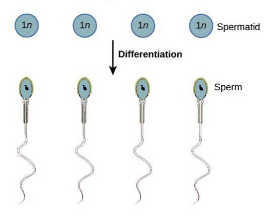
The spermatids of a mammal are characterized by having a rounded shape and lacking a flagellum, which is the whip-shaped appendix that helps movement, typical of sperm. Spermatids must mature into a sperm cell capable of performing its function: reaching the ovum and joining it.
Therefore, they must develop a flagellum morphologically reorganizing, thus acquiring motility and interaction capacity. The phases of spermiogenesis were described in 1963 and 1964 by Clermont and Heller, thanks to the visualization of each of the changes using light microcopy in human tissues..
The sperm differentiation process that occurs in mammals involves the following stages: the construction of an acrosomal vesicle, the formation of a hood, rotation and condensation of the nucleus.
Article index
Periodic acid granules, Schiff's reagent, abbreviated PAS, accumulate in the Golgi complex of spermatids..
PAS granules are rich in glycoproteins (carbohydrate-bound proteins) and will give rise to a vesicular structure called the acrosomal vesicle. During the Golgi phase, this vesicle increases in size.
The polarity of the sperm is defined by the position of the acrosomal vesicle and this structure will be located in the anterior pole of the sperm.
The acrosome is a structure that contains hydrolytic enzymes, such as hyaluronidase, trypsin and acrosin, whose function is the disintegration of the cells that accompany the oocyte, hydrolyzing the components of the matrix, such as hyaluronic acid..
This process is known as the acrosomal reaction and it begins with the contact between the sperm and the outermost layer of the oocyte, called the zona pellucida..
Another key event of the Golgi phase is the migration of the centrioles to the posterior region of the spermatid, and their alignment with the plasma membrane occurs..
The centriole proceeds to the assembly of the nine peripheral microtubules and the two central ones that make up the sperm flagellum..
This set of microtubules is capable of transforming energy - ATP (adenosine triphosphate) generated in the mitochondria - into movement..
The acrosomal vesicle proceeds to expand into the anterior half of the cell nucleus, giving the appearance of a helmet or cap. In this area, the nuclear envelope degenerates its pores and the structure thickens. In addition, condensation of the nucleus occurs.
During spermiogenesis, a series of transformations of the nucleus of the future sperm occurs, such as compaction to 10% of the initial size and the replacement of histones by protamines..
Protamines are proteins of about 5000 Da, rich in arginine, with less lysine, and soluble in water. These proteins are common in the sperm of different species and help in the extreme condemnation of DNA in an almost crystalline structure..
A change of orientation of the spermatid occurs: the head is arranged towards the Sertoli cells and the flagellum -in the process of development- extends inside the seminiferous tube.
The already condensed nucleus changes its shape, lengthening and taking on a more flattened shape. The nucleus, together with the acrosome, travels close to the plasma membrane at the anterior end.
In addition, a reorganization of the microtubules occurs in a cylindrical structure that widens from the acrosome to the posterior end of the spermatid..
As for the centrioles, after completing their function in the development of the flagellum, they return to the posterior area of the nucleus and adhere to it..
A series of modifications occurs to form the "neck" of the sperm. From the centrioles, now attached to the nucleus, emerge nine fibers of a significant diameter that spread in the tail outside the microtubules.
Note that these dense fibers join the nucleus with the flagellum; hence it is known as a "connecting piece".
The plasma membrane shifts to envelop the developing flagellum, and the mitochondria shift to form a helical structure around the neck that extends to the immediate posterior region.
The newly formed region is called the intermediate piece, located in the tail of the sperm. Likewise, the fibrous sheath, the main part and the main part can be distinguished.
The mitochondria originate a continuous covering that surrounds the intermediate piece, this layer is pyramid-shaped and participates in the generation of energy and in sperm movements.
The excess of cellular cytoplasmic content is phagocytosed by Sertoli cells, in the form of residual bodies.
After spermiogenesis, the sperm has radically changed its shape and is now a specialized cell with the ability to move.
In the spermatozoa generated, the head region can be differentiated (2-3 um wide and 4 to 5 um long), where the cell nucleus with the haploid genetic load and the acrosome are located.
After the head is the intermediate region, where the centrioles, the mitochondrial helix and the tail of about 50 um in length are located..
The spermiogenesis process varies depending on the species, although on average it lasts from one to three weeks. In experiments performed on mice, the sperm formation process takes 34.5 days. In contrast, the process in humans takes almost twice as long.
Spermatogenesis is a complete process that can occur continuously, generating about 100 million sperm per human testicle every day..
The release of sperm by ejaculation involves about 200 million. Throughout his life, a man can produce from 1012 up to 1013 sperm.


Yet No Comments