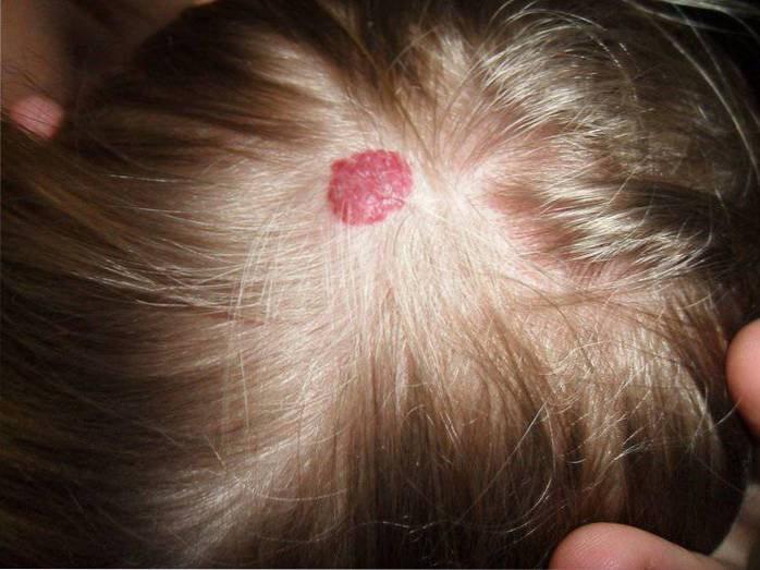
A brain hemangioma it is a type of vascular malformation characterized by clusters of dilated capillaries. They generally appear in brain and spinal areas, although it may occur on the retina or on the skin.
Brain hemangiomas can vary in size, from just a few millimeters to several centimeters in diameter, depending on the number of blood vessels involved. In some cases, those affected may have multiple lesions, while others will not experience a related clinical picture..
In the brain and spinal cord, these malformations, composed of very thin-walled capillaries, are very fragile and prone to bleeding, causing hemorrhagic strokes, seizures, and a wide variety of neurological deficits..
The signs and symptoms produced by this pathology will vary depending on the affected areas and secondary medical complications, however, some of the most frequent symptoms include muscle weakness or numbness, difficulty speaking, difficulty understanding others, headache. severe, sensory changes, instability, seizures, repeated bleeding, etc..
Brain hemangiomas are generally congenital in origin and brain imaging techniques are often used to identify their presence. In symptomatic cases, the treatment used is usually surgery, to eliminate the vascular malformation.
Article index
The Angioma Alliance points out that a cerebral hemangioma or cavernous angioma is an abnormal grouping of blood vessels at the brain, spinal level or in other areas of the body.
In addition, it points out that angiomas commonly have a structural configuration similar to a raspberry, composed of multiple bubbles (caverns), which contain blood inside and are covered with a thin layer of cells (endothelium)..
Due to both their shape and the lack of other supporting tissues, these blood vessels are prone to leaks and hemorrhages, giving rise to the development of the characteristic clinical picture of this pathology..
Although cavernous malformations can appear anywhere in the body, they usually only produce significant or more severe symptoms when they develop in the brain or spinal cord..
In addition, the clinical picture will vary depending on the number of vascular malformations, the location, the severity and the size. In many cases, these types of malformations can vary in size and number over time..
Hemangiomas or cavernous angiomas are a type of brain malformation that can occur in any age group and equally in men and women.
Statistical studies show that this pathology occurs in approximately 0.5-1% of the general population, that is, approximately 100-200 people.
Regarding the age of presentation of the first symptoms, it is frequent that the clinical course begins to develop between 20 and 30 years.
About 25% of those affected by cavernous malformations or cerebral hemangiomas do not usually experience significant signs or health problems related to this pathology.
However, in a good part of diagnosed cases, this medical condition can give rise to various serious signs and symptoms: convulsive episodes 30%, neurological deficit 25%, cerebral hemorrhage 15%, severe headache 5%.
Epileptic discharges are one of the most common symptoms of cavernous malformations. It is common for those affected to go to the emergency services and after control of the seizure episode, the presence of a cerebral hemangioma is discovered.
Approximately 30% of the cases of cavernous malformations will present seizures as one of the main symptoms..
Many of those affected may present various neurological alterations as a result of different brain and spinal cord injuries. The most common neurological disorders include double vision, muscle weakness, and even paralysis..
Generally the clinical symptoms are related to the place where the vascular malformation is located. Neurological deficits occur in approximately 25% of cerebral hemangioma cases.
15% of those affected by a cavernous angioma will present bleeding or cerebral hemorrhage. Specifically, brain hemorrhages are the most serious symptom of this type of pathology.
When bleeding begins, it is usually accompanied by a sudden headache followed by nausea, altered level of consciousness, or development of spontaneous neurological deficits.
In these cases, emergency medical care is essential since the life of the affected person is at serious risk if the volume of bleeding is high.
About 5% of people diagnosed with cerebral hemangioma go to informal suffering from severe headache of the headache or migraine type.
In relation to cerebral hemangiomas, two different forms of presentation of the pathology have been indicated: familial and sporadic.
It is a hereditary form of cerebral hemangiomas and transmission from father to son is frequent. Usually those affected tend to present multiple cavernous malformations at the brain level.
The familial form of cerebral hemangioma represents approximately 20% of all diagnosed cases and follows a dominant autonomic inheritance. The condition of this form has been associated with a genetic mutation in one of the following genes: CCM1, CCM2 or CCM3.
Specifically, mutations in the CCM3 gene lead to the development of the most severe form of cerebral hemangioma. Those affected are usually diagnosed at an early stage of life and present the first hemorrhages in childhood, they can also present cognitive alterations, benign brain tumors, skin lesions, etc..
Those affected by the sporadic form do not have a family history of the disease and usually present only an isolated brain malformation..
Experimental investigations have also identified genetic factors related to the development of the sporadic form of brain hemangiomas. Genetic mutations that are not heritable have been identified.
Therefore, people with an isolated cerebral cavernous malformation have a high probability of having the sporadic form, while people with multiple cavernous malformations have a higher probability of having the familial form..
As we have indicated previously, cerebral hemorrhages are the most serious and urgent symptom, since important transient or chronic neurological deficits can be derived from this..
Due to the absence of supporting tissues and the fragility of the capillaries that make up the cavernous malformation, they present a high probability of bleeding.
The Angioma Alliance notes that a cerebral hemangioma or cavernous angioma can bleed in different ways:
Bleeding may occur progressively and slowly within the walls of the brain angioma itself. Small-scale hemorrhages develop that do not usually require surgery, but their recurrence can lead to significant brain and spinal cord injuries..
It is also possible that bleeding occurs profusely within the walls of the brain angioma. Hemorrhages of a high magnitude develop that cause the size of the angioma to increase and exert pressure against the adjacent nerve tissues. It usually requires emergency medical intervention as it can generate significant neurological deficits.
Bleeding can break the walls of the angioma and therefore blood can reach the nerve tissues surrounding the angioma.
Although the risk of bleeding depends on the size and severity of the malformation, all cavernous angiomas have a high probability of bleeding.
It should be noted that up to approximately 40% of sporadically diagnosed cases of cerebral angiomas develop in parallel with another vascular anomaly, specifically venous angioma.
Venous angioma or developmental venous anomaly is a venous malformation in which a radial formation of veins can be observed that ends in a central or main one that is dilated. When it occurs in isolation, without cavernous angioma, it does not usually lead to the development of secondary medical complications (Angioma Alliance, 2016).
In addition to venous angiomas, cerebral hemangiomas can also develop associated with a type of lesion called "hidden vascular malformations", since they are not visible in some diagnostic tests such as angiograms..
When signs and symptoms compatible with the presence of a cavernous malformation are detected, there are two diagnostic tests that are usually used:
Both techniques are capable of providing images through brain sections and therefore allow medical specialists to locate the presence of a cerebral angioma..
Specifically, magnetic resonance imaging is capable of providing us with a view of hidden malformations in cerebral angiograms, providing high diagnostic precision..
Therefore, magnetic resonance imaging is the standard diagnostic technique in cavernous malformations, since these are not easily detected on computerized axial tomography or cerebral angiography..
On the other hand, the use of genetic tests makes it possible to identify genetic mutations related to familial and sporadic forms. Genetic tests are recommended for those affected with a family history of the disease or with multiple cavernous lesions.
In the therapeutic approach to cerebral hemangiomas, it is essential to take into account the following factors:
Therefore, depending on these factors, various approaches may be used, such as pharmacological, to control seizures and severe headache attacks. Apart from this, the fundamental treatment of cavernous angiomas is limited to surgical procedures..
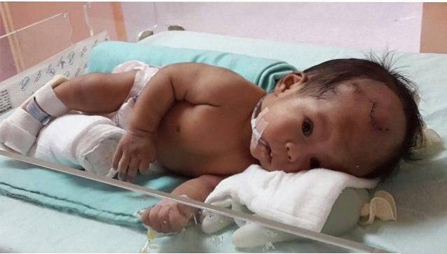
Brain hemangiomas are usually treated by surgical removal or resection through a craniotomy, or opening of the skull..
Despite the fact that this type of microsurgery is safe thanks to the technical advances that allow the lesion to be precisely located while minimally interrupting the rest of the functions, it also entails some risks such as: paralysis, coma and even death, although they are rare.
If the affected person undergoes a surgical resection procedure, they usually leave the hospital a few days after the intervention and regain a fully functional life.
Despite this, in cases where neurological dysfunction existed or develops later, it will be necessary to use physical and / or neuropsychological rehabilitation to recover altered functions or generate compensatory strategies.


Yet No Comments