Lymphoblast It is the name given to the immature precursor cells of two types of white blood cells (leukocytes) that circulate in the blood of mammalian animals: T lymphocytes and B lymphocytes; which are part of the group of immune cells.
In addition to these white blood cells, the cellular component of blood tissue is also represented by red blood cells (erythrocytes) and platelets (thrombocytes). While erythrocytes are responsible for the transport of oxygen through the body and platelets participate in clotting, leukocytes are part of the immune system.
Both red blood cells and white blood cells are formed through a process known as hemopoiesis, which occurs inside the bone marrow, from where they are released into the bloodstream, either fully mature, immature or partially mature, depending on the case.
Erythrocytes work directly in the circulatory system, but leukocytes use the bloodstream as a means of transport from one area of the body to another, where they exert their functions in the defense of the body against foreign substances or microorganisms.
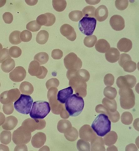
Lymphoblasts normally reside in the bone marrow, where they are produced; however, in some diseases such as lymphoblastic leukemia, lymphoblasts proliferate uncontrollably and can be found in large numbers in the peripheral blood, which means that they can be used as indicators that something is wrong.
Normally, the term "lymphoblast" is used to refer to immature forms of leukocytes, but there may be some inconsistencies in this regard in the literature..
Taking this definition into account, it is necessary to specify that they are precursor cells of two types of leukocytes in particular: T lymphocytes and B lymphocytes, which function, like most leukocytes, in the immune system.
It should also be clarified that white blood cells can be separated into two main categories:
Both groups of cells differ from each other with respect to the absence or presence in their interior of specific granules, respectively, which can be seen under the microscope and using a suitable staining method..
In the group of agranulocytes are monocytes and lymphocytes. Of these, lymphocytes represent approximately between 25 and 40% of the white blood cells found in the blood and are cells with the ability to migrate through the body's connective tissues..
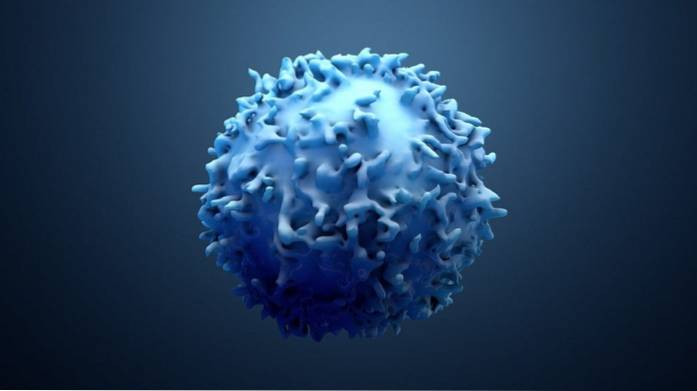
They are cells larger than erythrocytes and when observed in circulation they have a rounded appearance, while they can acquire different shapes - they are pleomorphic - when they enter or migrate towards the tissues.
The main tissues where these cells are concentrated are the spleen, tonsils, thymus and lymph nodes, which are primary lymphoid organs..
Lymphocytes have a nucleus with some depressions or notched edges that occupies most of the cytosolic space, which it shares with some small granules that are easy to differentiate under the microscope..
Although they are morphologically indistinguishable, according to the functions that these cells fulfill and some molecules that they present on their surface (markers), three types of lymphocytes are recognized:
Like T lymphocytes, B cells are fundamental cells for the adaptive immune system of mammalian animals and, in general terms, it is said that B lymphocytes are responsible for the humoral immune response and T lymphocytes for the cellular immune response..
These cells are capable of joining molecules derived from foreign substances or organisms, recognized as "not their own" -antigens- through a series of receptor molecules on their cell surface -antibodies- in order to eliminate them from the body or "Neutralize" them, preventing negative effects.
When they recognize a certain antigen, the lymphocytes multiply, thus producing cell clones capable of specifically recognizing that antigen, facilitating its rapid neutralization..
They also produce other cells known as memory cells, that form a kind of "library" of cells that recognize particular antigens and that can multiply when the body comes into contact with said antigens again, rapidly triggering the "educated" defense system.
Like all blood cells, lymphocytes derive from a type of resident cells of the bone marrow known as hematopoietic stem cells, which are capable of renewing and differentiating into different types of cells, that is, they are multipotent.
During the initial stages of hematopoiesis (production of red and white blood cells), two cell lines are established, which are derived from hematopoietic stem cells that differentiate into (1) lymphoid progenitor cells or (2) myeloid progenitor cells.
The differentiation of these stem cells implies that they are committed to a cell lineage, so they lose their ability to self-renew or produce new stem cells, as well as to differentiate into other types of cells.
When lymphocytes are produced in the bone marrow, they are actually immature cells (also called naive, virgins or not baited) that have not yet been exposed to any antigenic molecule, so they do not express on their surface any of the characteristic markers of this group of cells.
These immature cells are immunologically inactive, are approximately 6 microns in diameter, and remain in the G0 phase of the cell cycle. They have, in the cytosol, a series of rings around the nucleus, few mitochondria and poorly developed organelles.
When these immature lymphocytes interact with antigens, these molecules stimulate the advancement of their cell cycle from G0 to G1 and later to the synthesis phase, to the G2 phase and to the phase of mitosis and cell division..
Advancement in the cell cycle involves a series of internal transformations of the maturing lymphocytes, among which a considerable increase in size stands out (they can reach up to 15 microns)..
The maturing lymphocytes that have been stimulated by an antigen and that have increased in size are the "precursor" cells known as lymphoblasts..
These cells proliferate and end up differentiating into effector cells (antibody-producing cells: T lymphocytes and B lymphocytes) or in the memory cells that we talked about above..
Lymphoblasts have a well-defined nucleus within which a finely packed chromatin is evidenced. They usually have one or two nucleoli and a moderate amount of cytosol. These cells can actively divide, producing clones capable of recognizing the antigen that activated their proliferation..


Yet No Comments