The Valsalva maneuver It consists of changes in thoracic and abdominal pressure induced by forced expiration with the airways closed. The entire mechanism of this maneuver is completely voluntary and involves both times of breathing. Inspiration is followed by forced expiration opposed by a closed airway.
This maneuver owes its name to the Italian doctor Antonio Valsalva. In the seventeenth century the doctor studied the effects of expiration on the ear by keeping the mouth and nose covered. Valsalva was able to verify an opening of the Eustachian tube, communication between the middle ear and the pharynx; with this the pressure balance of the middle ear was achieved.
Sometimes the Valsalva maneuver occurs during daily activity; that is, the increase in pressure in the thoracoabdominal area. Lifting a heavy object, straining a bowel movement, sneezing, or coughing can produce this effect. Pushing is the common way of calling this maneuver.
Currently the Valsalva maneuver has many applications in the medical field. Diagnoses in cardiology, surgery, urology and neurosurgery are possible thanks to the use of this simple technique. Some therapeutic applications of the technique include achieving pressure compensation in the middle ear or reducing tachycardia..
Article index
The execution of the Valsalva maneuver involves the voluntary closure of the air outlet during a forced expiration. The occlusion of the airways is performed by covering the nose and mouth or causing a closure of the glottis. The purpose of the maneuver is to achieve an increase in pressure in both the thorax and the abdomen.
Once the increase in intrathoracic pressure occurs, a sequence of mechanisms occurs explained by the physiology of the maneuver. Just as in the thorax effects due to pressure are appreciated, in the abdominal organs they will also occur. Physiological changes during the Valsalva maneuver have been widely studied and described.
The physiological effect of the Valsalva maneuver within the thorax has been divided into four phases:
First, increased chest pressure causes increased pressure in the pulmonary veins. The pressure in the walls of the left atrium and ventricle will increase as a result of increased external pressure and blood flow..
The volume of blood leaving the heart increases, causing a transient rise in blood pressure.
By increasing the pressure within the thorax there is a drop in the volume of blood carried by the vena cava or venous return.
When this occurs, the volume of blood within the heart will be less, producing a decrease in cardiac output, which is directly proportional to venous return and heart rate..
The nervous system receives the signal of decreased cardiac output and generates a response through the autonomic nervous system. This response will be the release of adrenaline to produce an increase in heart rate, in compensation.
It is characterized by the recovery of cardiac output and a decrease in blood pressure. Once the intrathoracic pressure begins to decrease, the blood volume in the heart and vessels begins to balance. Heart rate and blood pressure decrease due to the regularization of cardiac output.
The cessation of the Valsalva maneuver determines the complete decrease in thoracic pressure. Venous return normalizes, allowing a volume of blood that was retained to enter the heart. Blood pressure will rise again due to sustained contraction of blood vessels.
The normal response at the end of the maneuver is the recovery of the physiological values of heart rate and blood pressure.
The diaphragm muscle anatomically divides the thoracic and abdominal cavities. The increase in pressure within the abdominal cavity will occur during the Valsalva maneuver as a consequence of the pressure exerted by the diaphragm. The muscles of the abdominal wall will also be contracted, contributing to the increase in pressure..
The great vessels, the abdominal and pelvic organs, and the spine will be affected as a result of increased intra-abdominal pressure..
Increased pressure on the inferior vena cava will decrease venous return from the lower limbs and abdominal organs.
The abdominal aorta will not be directly affected by changes in intra-abdominal pressure. Injuries to the aorta artery may be aggravated by the effect of the Valsalva.
The increase in peristalsis is an observed effect on the hollow viscera, in addition to the antegrade movement of its contents..
Pain due to inflammatory processes can be aggravated by the technique. The weaknesses of the abdominal wall will be evident during the execution of the maneuver.
The contraction of the abdominal and lumbar muscles, in addition to generating an increase in intra-abdominal pressure, will stabilize and strengthen the spine.
A similar effect is seen in the thoracic spine. Injuries at this level may be evidenced by pain from the pressure developed during the maneuver..
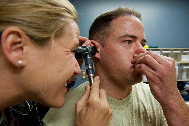
The Eustachian tube is a tube that connects the nasopharynx with the middle ear. Its function is to balance the pressure and drain the mucous secretion from that portion of the ear. The Eustachian tube contains air and remains closed.
Changes in atmospheric pressure can alter the pressure within the middle ear. This is commonly seen in divers or when traveling to high places. The Valsalva maneuver allows the Eustachian tube to open, thus balancing internal and external pressures.
The Valsalva maneuver currently has many applications in the medical field. The diagnostic value of this technique is greater than its therapeutic use.
It is a simple, non-instrumental technique that provides relevant data when performing a clinical examination. Its indication and proper execution do not involve health risks.
The cardiovascular physiological changes that occur during the Valsalva maneuver are useful both in the diagnosis and in the therapy of some diseases.
- Dilated cardiomyopathy or heart failure.
- Functional alteration of heart valves, such as aortic or pulmonary stenosis and mitral valve prolapse.
The therapeutic use of the Valsalva effect is limited to the correction of some arrhythmias, such as supraventricular tachycardia.
The diagnosis of abdominal wall weaknesses - such as hernias, hernias, or muscular diastasis - is achieved with the use of the Valsalva effect..
An increase in intra-abdominal pressure will reveal the existence of weak points in the abdomen. The use in urology can show the presence of varicoceles or disorders of the urinary system.
Acute surgical abdominal pain will prevent the performance of the Valsalva maneuver, since it will increase the pain produced by peritoneal irritation. In the postoperative period where spinal anesthesia has been used, the spinal fluid leak headache intensifies with the maneuver.
Compression of the nerve trunks exiting the spinal column produces pain or neurological symptoms. On occasions, during the physical examination, the patient is asked to perform the maneuver to reveal the presence of injuries, especially at the cervical or lumbar level..
The technique can also be useful in the physical examination after spinal interventions, such as laminectomies. Some headaches may get worse from this test..
- Labor is facilitated when intra-abdominal pressure is increased.
- For the diagnosis of genital prolapse.
- It is used for the diagnosis of the integrity of the hearing system.
- Evidence of sinusopathies.
- Balances middle ear pressure.
It is used to detect the existence of communication between the maxillary sinus and the oral cavity after a dental extraction.
Despite being a relatively simple diagnostic technique, the Valsalva maneuver should be used under surveillance and on medical advice. Contraindications to its use are due to the possibility of worsening some existing diseases in a person.
The Valsalva maneuver should not be performed in the following circumstances:
- Cardiovascular disorders, such as arrhythmias, high blood pressure, myocardial infarction, or aortic aneurysm.
- Suspicion of cerebrovascular disease, such as the presence of subarachnoid hemorrhage or aneurysms.
- Glaucoma.
- Tympanic rupture.
- Strangulated abdominal hernia.
- In pregnancy, when there is a threat of abortion or premature delivery.


Yet No Comments