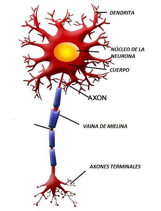
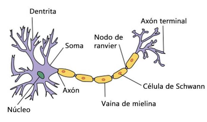
The myelin or myelin sheath is a fatty substance that surrounds nerve fibers and whose function is to increase the speed of nerve impulses, facilitating communication between neurons. It also allows greater energy savings for the nervous system.
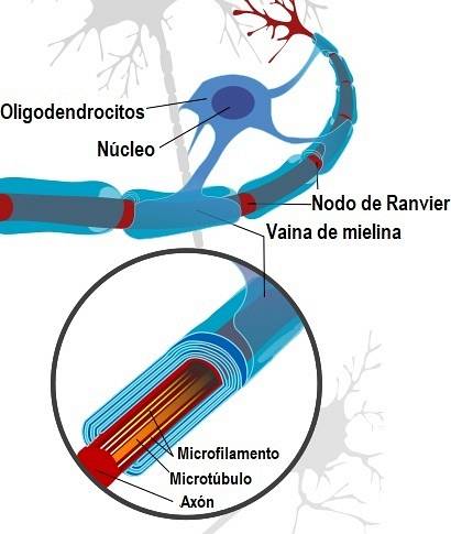
Myelin is made up of 80% lipids and 20% proteins. In the central nervous system, the nerve cells that produce it are glial cells called oligodendrocytes. While in the peripheral nervous system they are produced through Schwann cells.
The two main myelin proteins produced by oligodendrocytes are PLP (proteolipid protein) and MBP (myelin basic protein)..
When myelin doesn't develop properly or is injured for some reason, our nerve impulses slow down or become blocked. This is what happens in demyelinating diseases, leading to symptoms such as numbness, lack of coordination, paralysis, vision and cognitive problems.
Article index
This substance was discovered in the mid-1800s, but it took almost half a century before its important function as an insulator was revealed..
In the middle of the 19th century, scientists found something strange about the nerve fibers that branched out from the spinal cord. They observed that they were covered in a glistening white greasy substance.
The German pathologist Rudolf Virchow was the first to use the concept of "myelin." It comes from the Greek word "myelós", which means "marrow", referring to something central or internal.
This was because he thought that myelin was on the inside of nerve fibers. He incorrectly compared it to bone marrow.
Later, it was found that this substance enveloped the axons of neurons, forming sheaths. Regardless of where the myelin sheaths are located, the role is the same: efficiently transmit electrical signals.
In the 1870s, the French physician Louis-Antoine Ranvier noted that the myelin sheath is discontinuous. That is, there are gaps along the axon that do not have myelin. These have adopted the name of Ranvier's nodules, and serve to increase the speed of nerve conduction..
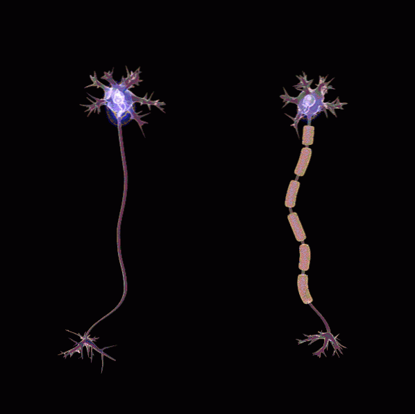
The myelin surrounds the axon or nerve extension forming a tube. The tube does not form a continuous covering, but is made up of a series of segments. Each of them is about 1mm.
Between the segments, there are small pieces of uncovered axon called Ranvier's nodules, measuring 1 to 2 micrometers..
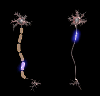
Thus, the myelin-coated axon resembles a string of elongated pearls. This facilitates the saltatory conduction of the nerve impulse, that is, the signals "jump" from one node to another. This allows the conduction speed to be faster in a myelinated neuron than in one without myelin..
Myelin also serves as an electrochemical insulator so that messages do not spread to adjacent cells and increases the resistance of the axon.
Beneath the cerebral cortex there are millions of axons that connect cortical neurons with those found in other parts of the brain. In this tissue there is a high concentration of myelin that gives it an opaque white color. Therefore, it is called white matter or white matter.
An oligodendrocyte can produce up to 50 servings of myelin. When the central nervous system is developing, these cells produce processes that resemble the oars of a canoe..
Then each of these are wound several times around a piece of axon, creating layers of myelin. Thanks to each paddle, therefore, a segment of the myelin sheath of an axon is obtained.
Myelin is also present in the peripheral nervous system, but it is produced by a type of nerve cells called Schwann cells..
Most of the axons in the peripheral nervous system are covered with myelin. The myelin sheaths are also segmented as in the central nervous system. Each myelinated area corresponds to a single Schwann cell that wraps itself several times around the axon..
The chemical composition of the myelin produced by oligodendrocytes and Schwann cells is different..
Therefore, in multiple sclerosis, the immune system of these patients only attacks the myelin protein produced by oligodendrocytes but not that generated by Schwann cells. Thus, the peripheral nervous system is not impaired.
All axons of the nervous systems of almost all mammals are covered with myelin sheaths. These are separated from each other by the nodules of Ranvier.
Action potentials travel differently through axons with myelin than through unmyelinated axons (lacking this substance).
The myelin coils around the axon without allowing extracellular fluid to penetrate between them. The only site on the axon that contacts the extracellular fluid is at the nodules of Ranvier, between each myelin sheath..
Thus, the action potential is produced and travels down the myelinated axon. As it travels through the myelin-filled area, the potential decreases, but it still has the strength to trigger another action potential in the next node. The potentials are repeated in each node of Ranvier, which is called "saltatory" conduction..
This type of conduction, facilitated by the structuring of myelin, allows impulses to travel much faster through our brain.
Thus, we can react in time to possible dangers, or develop cognitive tasks in seconds. In addition, this leads to great energy savings for our brain.
The myelination process is slow, beginning approximately 3 months after fertilization. It develops at different times depending on the area of the nervous system that is being formed.
For example, the prefrontal region is the last area to be myelinated, and it is the one in charge of complex functions such as planning, inhibition, motivation, self-regulation, etc..
At birth, only some areas of the brain are fully myelinated, such as the brain stem regions, which direct reflexes. Once their axons are myelinated, neurons achieve optimal functioning and faster and more efficient conduction.
Although the myelination process begins in an early postnatal period, the axons of the neurons of the cerebral hemispheres carry out this process a little later.
From the fourth month of life, neurons are myelinated until second childhood (between 6 and 12 years). It then continues through adolescence (12 to 18 years) through early adulthood, which is related to the development of complex cognitive functions.
The primary sensory and motor areas of the cerebral cortex begin their myelination before the frontal and parietal association zones. The latter are fully developed over 15 years.
The commissural, projection, and association fibers myelinate later than the primary sites. In fact, the structure that joins both cerebral hemispheres (called the corpus callosum), develops after birth and completes its myelination at 5 years. Greater myelination of the corpus callosum is associated with better cognitive functioning.
It has been proven that the myelination process goes in parallel with the cognitive development of the human being. The neuronal connections of the cerebral cortex become complex, and their myelination is related to the performance of increasingly elaborate behaviors.
For example, working memory has been shown to improve when the frontal lobe develops and myelinates. While the same occurs with visuospatial skills and myelination of the parietal area.
More complicated motor skills, such as sitting or walking, develop little by little in parallel with brain myelination..
The brain maturation process follows a vertical axis, starting in subcortical structures towards cortical structures (from the brain stem upwards). In addition, once inside the cortex, it maintains a horizontal direction, starting in the primary zones and continuing to the association regions..
This horizontal maturation leads to progressive changes within the same hemisphere of the brain. In addition, it establishes structural and functional differences between the two hemispheres..
Defective myelination is the main reason for neurological diseases. When axons lose their myelin, known as demyelination, nerve electrical signals are altered..
Demyelination can occur due to inflammation, metabolic or genetic problems. Whatever the cause, the loss of myelin causes significant nerve fiber dysfunction. Specifically, it reduces or blocks nerve impulses between the brain and the rest of the body..
Loss of myelin in humans has been linked to several central nervous system disorders such as stroke, spinal cord injury, and multiple sclerosis..
Some of the most common myelin-related diseases are:
In this disease, the immune system, which is responsible for defending the body from bacteria and viruses, mistakenly attacks the myelin sheaths. This causes the nerve cells and the spinal cord to be unable to communicate with each other or send messages to the muscles..
Symptoms range from fatigue, weakness, pain, and numbness, to paralysis and even vision loss. It also covers cognitive impairment and motor difficulties.
It appears due to a brief but intense inflammation of the brain and spinal cord that damages myelin. Vision loss, weakness, paralysis, and difficulty coordinating movements may occur.
Inflammation of the spinal cord that causes a loss of white matter in this place.
Other conditions include neuromyelitis optica, Guillain-Barré syndrome, or demyelinating polyneuropathies..
As for hereditary diseases that affect myelin, mention can be made of leukodystrophy and Charcot-Marie-Tooth disease. A more serious condition that severely damages myelin is Canavan disease..
The symptoms of demyelination are very diverse depending on the functions of the nerve cells involved. The manifestations vary according to each patient and disease, and have different clinical presentations according to each case. The most common symptoms are:
- Tiredness or fatigue.
- Vision problems: such as blurred vision in the center of the visual field, which affects only one eye. Pain may also appear when the eyes move. Another symptom is double vision or decreased vision..
- Hearing loss.
- Tinnitus or tinnitus, which is the perception of sounds or buzzing in the ears without external sources that produce them.
- Tingling or numbness in the legs, arms, face, or trunk. This is commonly known as neuropathy..
- Limb weakness.
- Symptoms worsen or reappear after exposure to heat, such as after a hot shower.
- Alteration of cognitive functions such as memory problems, or speech difficulties.
- Coordination, balance, or precision problems.
Myelin is currently being investigated to treat demyelinating diseases. Scientists seek to regenerate damaged myelin and prevent the chemical reactions that cause damage.
They are also developing drugs to stop or correct multiple sclerosis. In addition, they are investigating which specific antibodies are those that attack myelin and if stem cells could reverse the damage of demyelination.


Yet No Comments