The myxomycetes (Myxogastria class), also commonly known as plasmodia, slime molds or slime "fungi," are the most species-rich group within the phylum Amoebozoa, with approximately 1000 morphologically recognizable species. Due to the superficial resemblance of their reproductive structures they have been erroneously classified as fungi.
These organisms are unicellular protists without a cell wall, heterotrophs that feed on phagocytosis of bacteria, other protists, and fungi. They occupy diverse microhabitats in almost all terrestrial ecosystems and have even been located in aquatic environments. They live in the bark of trees, fallen or hanging plant debris and in the organic matter of the soil.
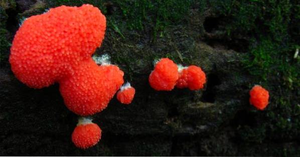
Specimens can be obtained as fruiting bodies grown under natural conditions or grown in the laboratory. The two trophic stages of their life cycle (amoeboflagellates and plasmodia) are often obscure, but the fruiting bodies are often large enough to be directly observed in nature..
They are not pathogenic, nor are they economically important. Only a few species are of interest as laboratory models; especially Physarum polycephalum Y Didymium iridis, have been used to investigate cell division and developmental biology in myxomycetes or to study some genetic mechanisms.
They fulfill a life cycle from spores generally spread through the air. They go through a haploid phase of flagellated uninucleated cells or not and a multinucleated diploid phase that ends in a fruiting body that gives rise to the sporrangia releasing the spores. They form resistance structures, microcysts and sclerotia, to survive extreme conditions.
Article index
General characteristics
Myxomycetes are unicellular, unicellular or plurinucleated, free-living terrestrial organisms, phagotrophic heterotrophs, lacking a cell wall. Spread by airborne spores or more rarely by animal vectors.
Since their discovery, myxomycetes have been variously classified as plants, animals, or fungi, because they produce aerial spores with structures that resemble those of certain fungi and typically occur in some of the same ecological situations as fungi..
The name Myxomycete, used for more than 175 years, is derived from the Greek words myxa (which means slime) and mycetes (referring to fungi).
However, the absence of a cell wall and their way of feeding by phagocytosis differentiate them from true fungi. Evidence obtained from RNA sequences confirms that they are amebozoans and not fungi.
Interestingly, the fact that Myxomycetes are protists was first pointed out more than a century and a half ago, when the name Mycetozoa was proposed for the group (literally meaning "animal mushroom")..
However, myxomycetos continued to be considered as fungi by most mycologists until the second half of the 20th century..
The first descriptions of organisms now known as Myxomycetes were supplied by Linnaeus in his Speies plantarum of 1753 (Lycoperdon epidendru, now called Lycogala epidendrum).
The first significant taxonomic treatment of the Myxomycetes was published by De Bary (1859), who was the first to conclude that these organisms were protists and not fungi..
The first monograph of the group is due to a De Bari student named Rostafinski (1873, 1874-1876). Because it was written in Polish, it was not widely circulated. The work that still remains as the definitive monograph for the group is The Myxomycetes, published by George Martin and Constantine Alexopoulos in 1969.
They belong to the supergroup Amoebozoa, in the class Myxogastria, and include two subclasses: Collumellidia and Lucisporidia. Due to the delicate nature of their structures, the fossil remains of Myxomycetes are not common, however some specimens of Stemonitis and Arcyria have been found in Baltic amber, dating back more than 50 million years. Phylogenetic studies with molecular data demonstrate its relationship with other groups of Amoebozoa and not with the kingdom Fungi.
Initially they were subdivided into six orders: Ceratiomyxales, Echinosteliales, Liceales, Physarales, Stemonitales, and Trichiales..
However, the members of the Ceratiomyxales, represented only by the genus Ceratiomyxa, they are clearly different from any of the organisms assigned to the other orders, hence they have been separated from the Myxomycetes.
For example, its spores are produced externally on individual stem structures and not within a fruiting body..
Recent molecular phylogenies have found a monophyletic clade (called "Macromycetozoan") composed of Dictyostelia, Myxogastria and Ceratiomyxa.
The Myxogastria group is monophyletic but deeply divided into two groups: the shiny spore Myxomycetes (Lucidisporidia) and the dark spore Myxomycetes (Columellidia). This difference is due to the appearance of melanin in the spore walls. Detailed phylogenetic relationships within the two groups have yet to be resolved.
60% of the known species have been detected directly in the field, recognizing their fruiting bodies, the other 40% are only known from being obtained in humid chambers or in agar culture media..
Myxomycetes are heterotrophs that feed on phagocytosis. Both in their form of ameboflagellates and plasmodia, their main food is free-living bacteria, but they also eat yeast, algae (including cyanobacteria) and fungi (spores and hyphae).
They are one of the most important groups in terms of bacterial consumption. Their location in the food chain assigns them an important ecological role by favoring the release of nutrients from the biomass of bacterial and fungal decomposers, especially vital nitrogen for plants..
They are widely distributed in almost all terrestrial ecosystems and some species even occupy aquatic habitats. An amoeboid organism related to the Myxomycetes has been isolated as endocomensal in the coelomic cavity of a sea urchin.
Temperature and humidity are the limiting factors for the occurrence of Myxomycetes in nature. In some cases the pH of the substrate can also influence.
They can inhabit extreme xeric conditions such as the Atacama Desert, parts of the Arabian Peninsula, the Gobi Desert in Mongolia or in alpine heights in the area where snow banks melt in late spring and early summer.
Their propagation and dormancy structures allow them to survive these extreme conditions: spores can survive for decades, microcysts and sclerotia for months or years..
The species richness of Myxomycetes tends to increase as the diversity and biomass of associated vegetation that gives rise to the debris that support the populations of bacteria and other microorganisms that serve as food increases. On the other hand, they adapt to very specific habitats, generating particular biotypes..
They are found growing on plant debris from the soil, tree bark (corticolas), living leaf surfaces (epiphiles), algae, hanging plant debris, inflorescences, manure from herbivorous animals.
The same species of Myxomycete will vary in color and size of the fruiting bodies depending on whether it develops in inflorescences of tropical herbs or in plant debris in the soil..
The Myxomycetes that usually appear on fallen trunks are the ones that generally produce larger fruiting bodies and for that reason they are the best known. This group includes species of the genera Arcyria, Lycogala, Stemonitis Y Trichia.
The life cycle of the Myxomycetes encompasses two very different trophic stages, one consisting of uninucleated amoebae, with or without flagella, and the other consisting of a distinctive multinucleate structure, the plasmodia, originated in most cases by sexual fusion. of the previous forms.
From the spore (haploid phase), a protoplast emerges. The protoplast can take the form of an amoeba capable of dividing or a non-divisible flagellate cell (the term amoeboflagellate refers to both forms).
These protoplasts divide by binary fission to build large populations in the various microhabitats where they develop. During the first trophic stage, in dry conditions or due to lack of food, an amoeboflagellate forms a microcyst or resting stage.
Compatible ameboflagellates form a zygote by gametic fusion, initiating the diploid phase. The nucleus of the zygote divides by mitosis and each new nucleus continues dividing without cytokinesis occurring, thus producing a single large multinucleated cell called plasmodia, which represents the second trophic phase..
Under adverse conditions, plasmodia can form the second type of resting structure found in myxomycetes: the sclerotia or macrocyst..
The entire plasmodium becomes a sporophor that generates fruiting bodies (also called sporocarps) that contain the spores formed by meiosis (haploid).
The spores of the Myxomycetes are dispersed by the wind or in some cases by animal vectors. An amoeboflagellate emerges from the spore and begins the cycle again.
However, some Myxomycetes are apomictic and do not exactly follow this cycle. Experiments carried out in monosporic cultures suggest that the colonies include a mixture of heterostallic (sexual) strains, where fusion of amoebae generates diploid plasmodia, and asexual strains where only ameboflagellates can mature into haploid plasmodia..


Yet No Comments